logo资料库

Deep Learning medical image analysis英文pdf.pdf

第1页 / 共459页
第2页 / 共459页
第3页 / 共459页
第4页 / 共459页
第5页 / 共459页
第6页 / 共459页
第7页 / 共459页
第8页 / 共459页
资料共459页,剩余部分请下载后查看
Front Cover
Deep Learning for Medical Image Analysis
Copyright
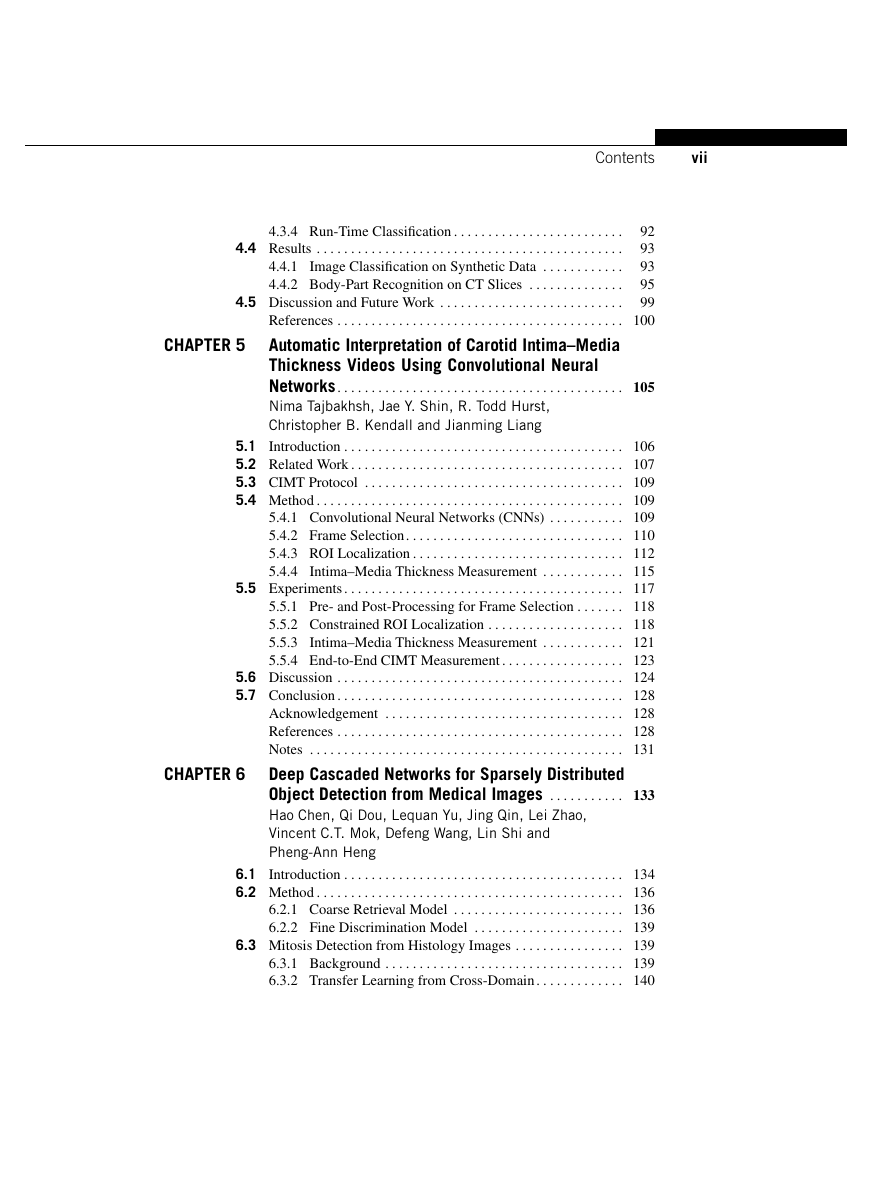
Contents
Contributors
About the Editors
Foreword
Part 1 Introduction
1 An Introduction to Neural Networks and Deep Learning
1.1 Introduction
1.2 Feed-Forward Neural Networks
1.2.1 Perceptron
1.2.2 Multi-Layer Neural Network
1.2.3 Learning in Feed-Forward Neural Networks
1.3 Convolutional Neural Networks
1.3.1 Convolution and Pooling Layer
1.3.2 Computing Gradients
1.4 Deep Models
1.4.1 Vanishing Gradient Problem
1.4.2 Deep Neural Networks
1.4.3 Deep Generative Models
1.5 Tricks for Better Learning
1.5.1 Rectified Linear Unit (ReLU)
1.5.2 Dropout
1.5.3 Batch Normalization
1.6 Open-Source Tools for Deep Learning
References
Notes
2 An Introduction to Deep Convolutional Neural Nets for Computer Vision
2.1 Introduction
2.2 Convolutional Neural Networks
2.2.1 Building Blocks of CNNs
2.2.2 Depth
2.2.3 Learning Algorithm
2.2.4 Tricks to Increase Performance
2.2.5 Putting It All Together: AlexNet
2.2.6 Using Pre-Trained CNNs
2.2.7 Improving AlexNet
2.3 CNN Flavors
2.3.1 Region-Based CNNs
2.3.2 Fully Convolutional Networks
2.3.3 Multi-Modal Networks
2.3.4 CNNs with RNNs
2.3.5 Hybrid Learning Methods
2.4 Software for Deep Learning
References
Part 2 Medical Image Detection and Recognition
3 Efficient Medical Image Parsing
3.1 Introduction
3.2 Background and Motivation
3.2.1 Object Localization and Segmentation: Challenges
3.3 Methodology
3.3.1 Problem Formulation
3.3.2 Sparse Adaptive Deep Neural Networks
3.3.3 Marginal Space Deep Learning
3.3.4 An Artificial Agent for Image Parsing
3.4 Experiments
3.4.1 Anatomy Detection and Segmentation in 3D
3.4.2 Landmark Detection in 2D and 3D
3.5 Conclusion
Disclaimer
References
4 Multi-Instance Multi-Stage Deep Learning for Medical Image Recognition
4.1 Introduction
4.2 Related Work
4.3 Methodology
4.3.1 Problem Statement and Framework Overview
4.3.2 Learning Stage I: Multi-Instance CNN Pre-Train
4.3.3 Learning Stage II: CNN Boosting
4.3.4 Run-Time Classification
4.4 Results
4.4.1 Image Classification on Synthetic Data
4.4.2 Body-Part Recognition on CT Slices
4.5 Discussion and Future Work
References
5 Automatic Interpretation of Carotid Intima-Media Thickness Videos Using Convolutional Neural Networks
5.1 Introduction
5.2 Related Work
5.3 CIMT Protocol
5.4 Method
5.4.1 Convolutional Neural Networks (CNNs)
5.4.2 Frame Selection
5.4.3 ROI Localization
5.4.4 Intima-Media Thickness Measurement
5.5 Experiments
5.5.1 Pre- and Post-Processing for Frame Selection
5.5.2 Constrained ROI Localization
5.5.3 Intima-Media Thickness Measurement
5.5.4 End-to-End CIMT Measurement
5.6 Discussion
5.7 Conclusion
Acknowledgement
References
Notes
6 Deep Cascaded Networks for Sparsely Distributed Object Detection from Medical Images
6.1 Introduction
6.2 Method
6.2.1 Coarse Retrieval Model
6.2.2 Fine Discrimination Model
6.3 Mitosis Detection from Histology Images
6.3.1 Background
6.3.2 Transfer Learning from Cross-Domain
6.3.3 Dataset and Preprocessing
6.3.4 Quantitative Evaluation and Comparison
6.3.5 Computation Cost
6.4 Cerebral Microbleed Detection from MR Volumes
6.4.1 Background
6.4.2 3D Cascaded Networks
6.4.3 Dataset and Preprocessing
6.4.4 Quantitative Evaluation and Comparison
6.4.5 System Implementation
6.5 Discussion and Conclusion
Acknowledgements
References
Notes
7 Deep Voting and Structured Regression for Microscopy Image Analysis
7.1 Deep Voting: A Robust Approach Toward Nucleus Localization in Microscopy Images
7.1.1 Introduction
7.1.2 Methodology
7.1.3 Weighted Voting Density Estimation
7.1.4 Experiments
7.1.5 Conclusion
7.2 Structured Regression for Robust Cell Detection Using Convolutional Neural Network
7.2.1 Introduction
7.2.2 Methodology
7.2.3 Experimental Results
7.2.4 Conclusion
Acknowledgements
References
Part 3 Medical Image Segmentation
8 Deep Learning Tissue Segmentation in Cardiac Histopathology Images
8.1 Introduction
8.2 Experimental Design and Implementation
8.2.1 Data Set Description
8.2.2 Manual Ground Truth Annotations
8.2.3 Implementation
8.2.4 Training a Model Using Engineered Features
8.2.5 Experiments
8.2.6 Testing and Performance Evaluation
8.3 Results and Discussion
8.3.1 Experiment 1: Comparison of Deep Learning and Random Forest Segmentation
8.3.2 Experiment 2: Evaluating the Sensitivity of Deep Learning to Training Data
8.4 Concluding Remarks
Notes
Disclosure Statement
Funding
References
9 Deformable MR Prostate Segmentation via Deep Feature Learning and Sparse Patch Matching
9.1 Background
9.2 Proposed Method
9.2.1 Related Work
9.2.2 Learning Deep Feature Representation
9.2.3 Segmentation Using Learned Feature Representation
9.3 Experiments
9.3.1 Evaluation of the Performance of Deep-Learned Features
9.3.2 Evaluation of the Performance of Deformable Model
9.4 Conclusion
References
10 Characterization of Errors in Deep Learning-Based Brain MRI Segmentation
10.1 Introduction
10.2 Deep Learning for Segmentation
10.3 Convolutional Neural Network Architecture
10.3.1 Basic CNN Architecture
10.3.2 Tri-Planar CNN for 3D Image Analysis
10.4 Experiments
10.4.1 Dataset
10.4.2 CNN Parameters
10.4.3 Training
10.4.4 Estimation of Centroid Distances
10.4.5 Registration-Based Segmentation
10.4.6 Characterization of Errors
10.5 Results
10.5.1 Overall Performance
10.5.2 Errors
10.6 Discussion
10.7 Conclusion
References
Notes
Part 4 Medical Image Registration
11 Scalable High Performance Image Registration Framework by Unsupervised Deep Feature Representations Learning
11.1 Introduction
11.2 Proposed Method
11.2.1 Related Research
11.2.2 Learn Intrinsic Feature Representations by Unsupervised Deep Learning
11.2.3 Registration Using Learned Feature Representations
11.3 Experiments
11.3.1 Experimental Result on ADNI Dataset
11.3.2 Experimental Result on LONI Dataset
11.3.3 Experimental Result on 7.0-T MR Image Dataset
11.4 Conclusion
References
12 Convolutional Neural Networks for Robust and Real-Time 2-D/3-D Registration
12.1 Introduction
12.2 X-Ray Imaging Model
12.3 Problem Formulation
12.4 Regression Strategy
12.4.1 Parameter Space Partitioning
12.4.2 Marginal Space Regression
12.5 Feature Extraction
12.5.1 Local Image Residual
12.5.2 3-D Points of Interest
12.6 Convolutional Neural Network
12.6.1 Network Structure
12.6.2 Training Data
12.6.3 Solver
12.7 Experiments and Results
12.7.1 Experiment Setup
12.7.2 Hardware & Software
12.7.3 Performance Analysis
12.7.4 Comparison with State-of-the-Art Methods
12.8 Discussion
Disclaimer
References
Part 5 Computer-Aided Diagnosis and Disease Quantification
13 Chest Radiograph Pathology Categorization via Transfer Learning
13.1 Introduction
13.2 Image Representation Schemes with Classical (Non-Deep) Features
13.2.1 Classical Filtering
13.2.2 Bag-of-Visual-Words Model
13.3 Extracting Deep Features from a Pre-Trained CNN Model
13.4 Extending the Representation Using Feature Fusion and Selection
13.5 Experiments and Results
13.5.1 Data
13.5.2 Experimental Setup
13.5.3 Experimental Results
13.6 Conclusion
Acknowledgements
References
14 Deep Learning Models for Classifying Mammogram Exams Containing Unregistered Multi-View Images and Segmentation Maps of Lesions
14.1 Introduction
14.2 Literature Review
14.3 Methodology
14.3.1 Deep Learning Model
14.4 Materials and Methods
14.5 Results
14.6 Discussion
14.7 Conclusion
Acknowledgements
References
Note
15 Randomized Deep Learning Methods for Clinical Trial Enrichment and Design in Alzheimer's Disease
15.1 Introduction
15.2 Background
15.2.1 Clinical Trials and Sample Enrichment
15.2.2 Neural Networks
15.2.3 Backpropagation and Deep Learning
15.3 Optimal Enrichment Criterion
15.3.1 Ensemble Learning and Randomization
15.4 Randomized Deep Networks
15.4.1 Small Sample Regime and Multiple Modalities
15.4.2 Roadmap
15.4.3 RDA and RDR Training
15.4.4 The Disease Markers - RDAM and RDRM
15.5 Experiments
15.5.1 Participant Data and Preprocessing
15.5.2 Evaluations Setup
15.5.3 Results
15.6 Discussion
Acknowledgements
References
Notes
Part 6 Others
16 Deep Networks and Mutual Information Maximization for Cross-Modal Medical Image Synthesis
16.1 Introduction
16.2 Supervised Synthesis Using Location-Sensitive Deep Network
16.2.1 Backpropagation
16.2.2 Network Simplification
16.2.3 Experiments
16.3 Unsupervised Synthesis Using Mutual Information Maximization
16.3.1 Generating Multiple Target Modality Candidates
16.3.2 Full Image Synthesis Using Best Candidates
16.3.3 Refinement Using Coupled Sparse Representation
16.3.4 Extension to Supervised Setting
16.3.5 Experiments
16.4 Conclusions and Future Work
Acknowledgements
References
Note
17 Natural Language Processing for Large-Scale Medical Image Analysis Using Deep Learning
17.1 Introduction
17.2 Fundamentals of Natural Language Processing
17.2.1 Pattern Matching
17.2.2 Topic Modeling
17.3 Neural Language Models
17.3.1 Word Embeddings
17.3.2 Recurrent Language Model
17.4 Medical Lexicons
17.4.1 UMLS Metathesaurus
17.4.2 RadLex
17.5 Predicting Presence or Absence of Frequent Disease Types
17.5.1 Mining Presence/Absence of Frequent Disease Terms
17.5.2 Prediction Result and Discussion
17.6 Conclusion
Acknowledgements
References
Index
Back Cover
Deep Learning for Medical Image Analysis Edited by S. Kevin Zhou Hayit Greenspan Dinggang Shen
Deep Learning for Medical Image Analysis
The Elsevier and MICCAI Society Book Series Advisory board Stephen Aylward (Kitware,UnitedStates) David Hawkes (UniversityCollegeLondon,UnitedKingdom) Kensaku Mori (UniversityofNagoya,Japan) Alison Noble (UniversityofOxford,UnitedKingdom) Sonia Pujol (HarvardUniversity,UnitedStates) Daniel Rueckert (ImperialCollege,UnitedKingdom) Xavier Pennec (INRIASophia-Antipolis,France) Pierre Jannin (UniversityofRennes,France) Also available: Balocco, Computing and Visualization for Intravascular Imaging and Computer Assisted Stenting, 9780128110188 Wu, Machine Learning and Medical Imaging, 9780128040768 Zhou, Medical Image Recognition, Segmentation and Parsing, 9780128025819
Deep Learning for Medical Image Analysis Edited by S. Kevin Zhou Hayit Greenspan Dinggang Shen
Academic Press is an imprint of Elsevier 125 London Wall, London EC2Y 5AS, United Kingdom 525 B Street, Suite 1800, San Diego, CA 92101-4495, United States 50 Hampshire Street, 5th Floor, Cambridge, MA 02139, United States The Boulevard, Langford Lane, Kidlington, Oxford OX5 1GB, United Kingdom Copyright © 2017 Elsevier Inc. All rights reserved. No part of this publication may be reproduced or transmitted in any form or by any means, electronic or mechanical, including photocopying, recording, or any information storage and retrieval system, without permission in writing from the publisher. Details on how to seek permission, further information about the Publisher’s permissions policies and our arrangements with organizations such as the Copyright Clearance Center and the Copyright Licensing Agency, can be found at our website: www.elsevier.com/permissions. This book and the individual contributions contained in it are protected under copyright by the Publisher (other than as may be noted herein). Notices Knowledge and best practice in this field are constantly changing. As new research and experience broaden our understanding, changes in research methods, professional practices, or medical treatment may become necessary. Practitioners and researchers must always rely on their own experience and knowledge in evaluating and using any information, methods, compounds, or experiments described herein. In using such information or methods they should be mindful of their own safety and the safety of others, including parties for whom they have a professional responsibility. To the fullest extent of the law, neither the Publisher nor the authors, contributors, or editors, assume any liability for any injury and/or damage to persons or property as a matter of products liability, negligence or otherwise, or from any use or operation of any methods, products, instructions, or ideas contained in the material herein. Library of Congress Cataloging-in-Publication Data A catalog record for this book is available from the Library of Congress British Library Cataloguing-in-Publication Data A catalogue record for this book is available from the British Library ISBN: 978-0-12-810408-8 For information on all Academic Press publications visit our website at https://www.elsevier.com Publisher: Joe Hayton Acquisition Editor: Tim Pitts Editorial Project Manager: Charlotte Kent Production Project Manager: Julie-Ann Stansfield Designer: Matthew Limbert Typeset by VTeX
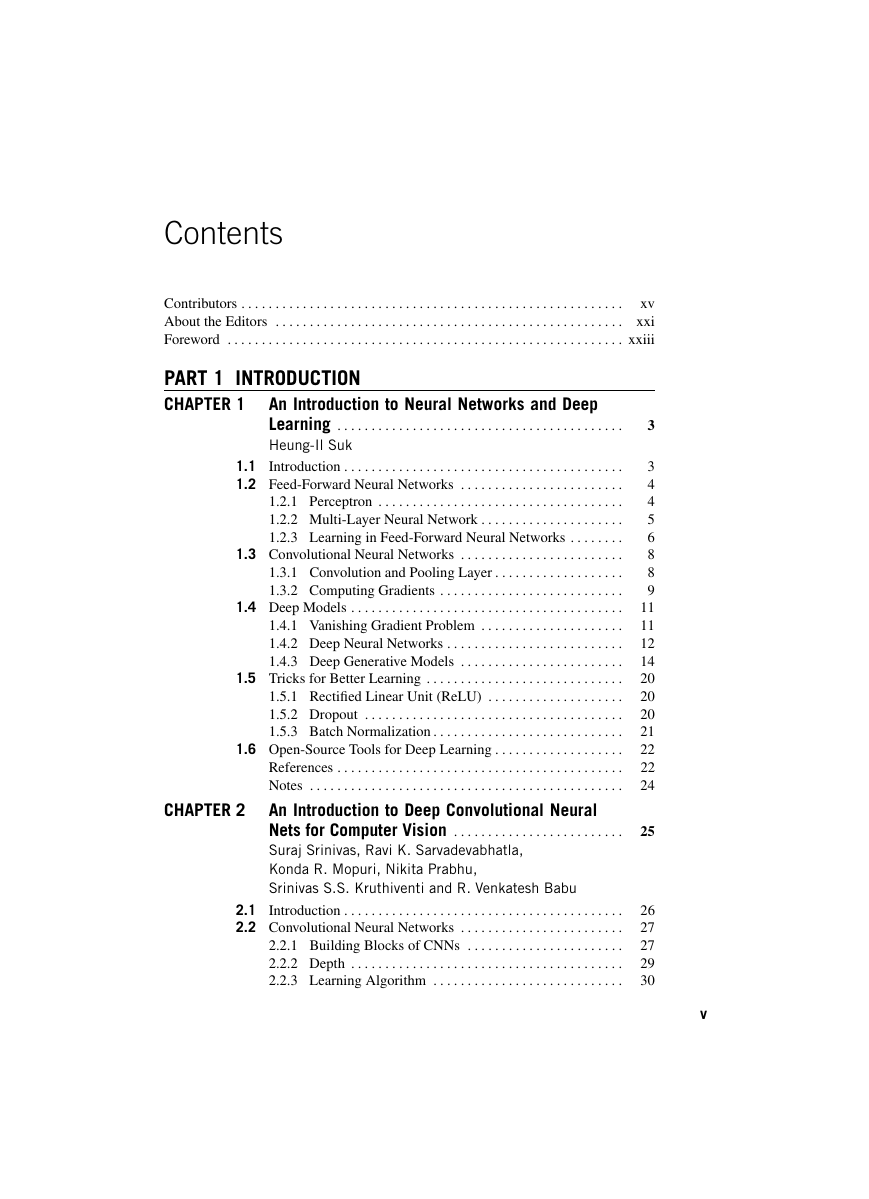
Contents xv Contributors . . . . . . . . . . . . . . . . . . . . . . . . . . . . . . . . . . . . . . . . . . . . . . . . . . . . . . . . About the Editors . . . . . . . . . . . . . . . . . . . . . . . . . . . . . . . . . . . . . . . . . . . . . . . . . . . xxi Foreword . . . . . . . . . . . . . . . . . . . . . . . . . . . . . . . . . . . . . . . . . . . . . . . . . . . . . . . . . . xxiii PART 1 INTRODUCTION CHAPTER 1 An Introduction to Neural Networks and Deep Learning . . . . . . . . . . . . . . . . . . . . . . . . . . . . . . . . . . . . . . . . . . Heung-Il Suk 1.1 Introduction . . . . . . . . . . . . . . . . . . . . . . . . . . . . . . . . . . . . . . . . . 1.2 Feed-Forward Neural Networks . . . . . . . . . . . . . . . . . . . . . . . . 1.2.1 Perceptron . . . . . . . . . . . . . . . . . . . . . . . . . . . . . . . . . . . . 1.2.2 Multi-Layer Neural Network . . . . . . . . . . . . . . . . . . . . . 1.2.3 Learning in Feed-Forward Neural Networks . . . . . . . . 1.3 Convolutional Neural Networks . . . . . . . . . . . . . . . . . . . . . . . . 1.3.1 Convolution and Pooling Layer . . . . . . . . . . . . . . . . . . . 1.3.2 Computing Gradients . . . . . . . . . . . . . . . . . . . . . . . . . . . 1.4 Deep Models . . . . . . . . . . . . . . . . . . . . . . . . . . . . . . . . . . . . . . . . 1.4.1 Vanishing Gradient Problem . . . . . . . . . . . . . . . . . . . . . 1.4.2 Deep Neural Networks . . . . . . . . . . . . . . . . . . . . . . . . . . 1.4.3 Deep Generative Models . . . . . . . . . . . . . . . . . . . . . . . . 1.5 Tricks for Better Learning . . . . . . . . . . . . . . . . . . . . . . . . . . . . . 1.5.1 Rectified Linear Unit (ReLU) . . . . . . . . . . . . . . . . . . . . 1.5.2 Dropout . . . . . . . . . . . . . . . . . . . . . . . . . . . . . . . . . . . . . . 1.5.3 Batch Normalization . . . . . . . . . . . . . . . . . . . . . . . . . . . . 1.6 Open-Source Tools for Deep Learning . . . . . . . . . . . . . . . . . . . References . . . . . . . . . . . . . . . . . . . . . . . . . . . . . . . . . . . . . . . . . . Notes . . . . . . . . . . . . . . . . . . . . . . . . . . . . . . . . . . . . . . . . . . . . . . An Introduction to Deep Convolutional Neural Nets for Computer Vision . . . . . . . . . . . . . . . . . . . . . . . . . Suraj Srinivas, Ravi K. Sarvadevabhatla, Konda R. Mopuri, Nikita Prabhu, Srinivas S.S. Kruthiventi and R. Venkatesh Babu CHAPTER 2 2.1 Introduction . . . . . . . . . . . . . . . . . . . . . . . . . . . . . . . . . . . . . . . . . 2.2 Convolutional Neural Networks . . . . . . . . . . . . . . . . . . . . . . . . 2.2.1 Building Blocks of CNNs . . . . . . . . . . . . . . . . . . . . . . . 2.2.2 Depth . . . . . . . . . . . . . . . . . . . . . . . . . . . . . . . . . . . . . . . . 2.2.3 Learning Algorithm . . . . . . . . . . . . . . . . . . . . . . . . . . . . 3 3 4 4 5 6 8 8 9 11 11 12 14 20 20 20 21 22 22 24 25 26 27 27 29 30 v
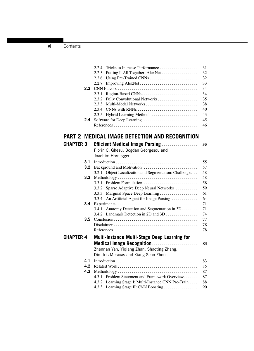
vi Contents 2.2.4 Tricks to Increase Performance . . . . . . . . . . . . . . . . . . . 2.2.5 Putting It All Together: AlexNet . . . . . . . . . . . . . . . . . . 2.2.6 Using Pre-Trained CNNs . . . . . . . . . . . . . . . . . . . . . . . . 2.2.7 Improving AlexNet . . . . . . . . . . . . . . . . . . . . . . . . . . . . . 2.3 CNN Flavors . . . . . . . . . . . . . . . . . . . . . . . . . . . . . . . . . . . . . . . . 2.3.1 Region-Based CNNs . . . . . . . . . . . . . . . . . . . . . . . . . . . . 2.3.2 Fully Convolutional Networks . . . . . . . . . . . . . . . . . . . . 2.3.3 Multi-Modal Networks . . . . . . . . . . . . . . . . . . . . . . . . . . 2.3.4 CNNs with RNNs . . . . . . . . . . . . . . . . . . . . . . . . . . . . . . 2.3.5 Hybrid Learning Methods . . . . . . . . . . . . . . . . . . . . . . . 2.4 Software for Deep Learning . . . . . . . . . . . . . . . . . . . . . . . . . . . References . . . . . . . . . . . . . . . . . . . . . . . . . . . . . . . . . . . . . . . . . . PART 2 MEDICAL IMAGE DETECTION AND RECOGNITION Efficient Medical Image Parsing . . . . . . . . . . . . . . . . . . CHAPTER 3 Florin C. Ghesu, Bogdan Georgescu and Joachim Hornegger 3.1 Introduction . . . . . . . . . . . . . . . . . . . . . . . . . . . . . . . . . . . . . . . . . 3.2 Background and Motivation . . . . . . . . . . . . . . . . . . . . . . . . . . . 3.2.1 Object Localization and Segmentation: Challenges . . 3.3 Methodology . . . . . . . . . . . . . . . . . . . . . . . . . . . . . . . . . . . . . . . . 3.3.1 Problem Formulation . . . . . . . . . . . . . . . . . . . . . . . . . . . 3.3.2 Sparse Adaptive Deep Neural Networks . . . . . . . . . . . 3.3.3 Marginal Space Deep Learning . . . . . . . . . . . . . . . . . . . 3.3.4 An Artificial Agent for Image Parsing . . . . . . . . . . . . . 3.4 Experiments . . . . . . . . . . . . . . . . . . . . . . . . . . . . . . . . . . . . . . . . . 3.4.1 Anatomy Detection and Segmentation in 3D . . . . . . . . 3.4.2 Landmark Detection in 2D and 3D . . . . . . . . . . . . . . . . 3.5 Conclusion . . . . . . . . . . . . . . . . . . . . . . . . . . . . . . . . . . . . . . . . . . Disclaimer . . . . . . . . . . . . . . . . . . . . . . . . . . . . . . . . . . . . . . . . . . References . . . . . . . . . . . . . . . . . . . . . . . . . . . . . . . . . . . . . . . . . . CHAPTER 4 Multi-Instance Multi-Stage Deep Learning for Medical Image Recognition . . . . . . . . . . . . . . . . . . . . . . . Zhennan Yan, Yiqiang Zhan, Shaoting Zhang, Dimitris Metaxas and Xiang Sean Zhou 4.1 Introduction . . . . . . . . . . . . . . . . . . . . . . . . . . . . . . . . . . . . . . . . . 4.2 Related Work . . . . . . . . . . . . . . . . . . . . . . . . . . . . . . . . . . . . . . . . 4.3 Methodology . . . . . . . . . . . . . . . . . . . . . . . . . . . . . . . . . . . . . . . . 4.3.1 Problem Statement and Framework Overview . . . . . . . 4.3.2 Learning Stage I: Multi-Instance CNN Pre-Train . . . . 4.3.3 Learning Stage II: CNN Boosting . . . . . . . . . . . . . . . . . 31 32 32 33 34 34 35 38 40 43 45 46 55 55 57 58 58 58 59 61 64 71 71 74 77 78 78 83 83 85 87 87 88 90
Contents vii 4.3.4 Run-Time Classification . . . . . . . . . . . . . . . . . . . . . . . . . 4.4 Results . . . . . . . . . . . . . . . . . . . . . . . . . . . . . . . . . . . . . . . . . . . . . 4.4.1 Image Classification on Synthetic Data . . . . . . . . . . . . 4.4.2 Body-Part Recognition on CT Slices . . . . . . . . . . . . . . 4.5 Discussion and Future Work . . . . . . . . . . . . . . . . . . . . . . . . . . . 92 93 93 95 99 References . . . . . . . . . . . . . . . . . . . . . . . . . . . . . . . . . . . . . . . . . . 100 Automatic Interpretation of Carotid Intima–Media Thickness Videos Using Convolutional Neural Networks . . . . . . . . . . . . . . . . . . . . . . . . . . . . . . . . . . . . . . . . . . 105 Nima Tajbakhsh, Jae Y. Shin, R. Todd Hurst, Christopher B. Kendall and Jianming Liang CHAPTER 5 5.1 Introduction . . . . . . . . . . . . . . . . . . . . . . . . . . . . . . . . . . . . . . . . . 106 5.2 Related Work . . . . . . . . . . . . . . . . . . . . . . . . . . . . . . . . . . . . . . . . 107 5.3 CIMT Protocol . . . . . . . . . . . . . . . . . . . . . . . . . . . . . . . . . . . . . . 109 5.4 Method . . . . . . . . . . . . . . . . . . . . . . . . . . . . . . . . . . . . . . . . . . . . . 109 5.4.1 Convolutional Neural Networks (CNNs) . . . . . . . . . . . 109 5.4.2 Frame Selection . . . . . . . . . . . . . . . . . . . . . . . . . . . . . . . . 110 5.4.3 ROI Localization . . . . . . . . . . . . . . . . . . . . . . . . . . . . . . . 112 5.4.4 Intima–Media Thickness Measurement . . . . . . . . . . . . 115 5.5 Experiments . . . . . . . . . . . . . . . . . . . . . . . . . . . . . . . . . . . . . . . . . 117 5.5.1 Pre- and Post-Processing for Frame Selection . . . . . . . 118 5.5.2 Constrained ROI Localization . . . . . . . . . . . . . . . . . . . . 118 5.5.3 Intima–Media Thickness Measurement . . . . . . . . . . . . 121 5.5.4 End-to-End CIMT Measurement . . . . . . . . . . . . . . . . . . 123 5.6 Discussion . . . . . . . . . . . . . . . . . . . . . . . . . . . . . . . . . . . . . . . . . . 124 5.7 Conclusion . . . . . . . . . . . . . . . . . . . . . . . . . . . . . . . . . . . . . . . . . . 128 Acknowledgement . . . . . . . . . . . . . . . . . . . . . . . . . . . . . . . . . . . 128 References . . . . . . . . . . . . . . . . . . . . . . . . . . . . . . . . . . . . . . . . . . 128 Notes . . . . . . . . . . . . . . . . . . . . . . . . . . . . . . . . . . . . . . . . . . . . . . 131 Deep Cascaded Networks for Sparsely Distributed Object Detection from Medical Images . . . . . . . . . . . 133 Hao Chen, Qi Dou, Lequan Yu, Jing Qin, Lei Zhao, Vincent C.T. Mok, Defeng Wang, Lin Shi and Pheng-Ann Heng CHAPTER 6 6.1 Introduction . . . . . . . . . . . . . . . . . . . . . . . . . . . . . . . . . . . . . . . . . 134 6.2 Method . . . . . . . . . . . . . . . . . . . . . . . . . . . . . . . . . . . . . . . . . . . . . 136 6.2.1 Coarse Retrieval Model . . . . . . . . . . . . . . . . . . . . . . . . . 136 6.2.2 Fine Discrimination Model . . . . . . . . . . . . . . . . . . . . . . 139 6.3 Mitosis Detection from Histology Images . . . . . . . . . . . . . . . . 139 6.3.1 Background . . . . . . . . . . . . . . . . . . . . . . . . . . . . . . . . . . . 139 6.3.2 Transfer Learning from Cross-Domain . . . . . . . . . . . . . 140
分享到:
收藏