Preface
Organization
Contents – Part III
Diffusion Tensor Imaging and Functional MRI: Diffusion Tensor Imaging
Multimodal Fusion of Brain Networks with Longitudinal Couplings
1 Introduction
2 Brian Network Representation Learning
2.1 Modeling Cross-Sectional Coupling
2.2 Modeling Multimodal Coupling
2.3 Modeling Longitudinal Coupling
2.4 The Proposed Model - MMLC
3 An Optimization Framework for MMLC
4 Experiment
4.1 Experimental Settings and Baseline Methods
4.2 Results
5 Conclusion and Future Works
References
Penalized Geodesic Tractography for Mitigating Gyral Bias
1 Introduction
2 Methods
2.1 Geodesic Tractography
2.2 Penalized Finsler Metric
2.3 Tractography with Optimized Shortest Path
2.4 Datasets and Processing
3 Results
4 Conclusion
References
Anchor-Constrained Plausibility (ACP): A Novel Concept for Assessing Tractography and Reducing False-Positives
1 Introduction
2 Methods
3 Experiments and Results
3.1 In Silico Experiments and Results
3.2 In Vivo Experiments and Results
4 Discussion and Conclusion
References
Tract-Specific Group Analysis in Fetal Cohorts Using in utero Diffusion Tensor Imaging
1 Introduction
2 Methods
2.1 DWI Processing and DTI Computation
2.2 Study-Specific Template and Normalization
2.3 Statistical Analysis
3 Application and Results
4 Conclusion
References
Tract Orientation Mapping for Bundle-Specific Tractography
1 Introduction
2 Methods
3 Results
4 Discussion and Conclusion
References
A Multi-Tissue Global Estimation Framework for Asymmetric Fiber Orientation Distributions
1 Introduction
2 Method
2.1 Asymmetric Fiber Orientation Distribution
2.2 Global Estimation Framework
2.3 Optimization
2.4 Materials and Implementation Details
3 Experimental Results
4 Conclusion
References
Diffusion Tensor Imaging and Functional MRI: Diffusion Weighted Imaging
Better Fiber ODFs from Suboptimal Data with Autoencoder Based Regularization
1 Introduction
2 Background and Related Work
3 Hybrid fODF Estimation
3.1 Role and Training of Autoencoders in Our Method
3.2 Computational Pipeline and Theoretical Guarantees
4 Results and Discussion
4.1 Quantitative Results
4.2 Practical Benefit from Theoretical Guarantees
4.3 Iterating Our Method
4.4 Qualitative Results
5 Conclusion
References
Identification of Gadolinium Contrast Enhanced Regions in MS Lesions Using Brain Tissue Microstructure Information Obtained from Diffusion and T2 Relaxometry MRI
1 Introduction
2 Materials and Methods
2.1 Multi-Compartment T2 Relaxometry Model (MCT2)
2.2 Multi-Compartment Diffusion Model (MCDiff)
2.3 Data
2.4 Identifying Enhancing Voxels in Lesions
3 Results
4 Discussion
5 Conclusion
References
A Bayes Hilbert Space for Compartment Model Computing in Diffusion MRI
1 Introduction
2 Theory
2.1 Bayes Hilbert Spaces and Compartmental Diffusion
2.2 Gaussian Compartmental Diffusion
3 Experimental Setup
3.1 Simulations: Robustness to Noise
3.2 Case Study: Tractography of the Pyramidal Tract
4 Results and Discussion
References
Detection and Delineation of Acute Cerebral Infarct on DWI Using Weakly Supervised Machine Learning
1 Introduction
2 Data
2.1 Weak Labels
2.2 Manual Segmentation
2.3 Imaging Data
3 Model
3.1 Semi-Supervised Learning
4 Experiments and Results
5 Discussion and Conclusion
References
Identification of Species-Preserved Cortical Landmarks
Abstract
1 Introduction
2 Materials and Methods
2.1 Overview
2.2 Data Set Description and Pre-processing
2.3 Identification of 3-hinges and Reconstruction of Connective Matrix
2.4 Identification of Consistent 3-hinge Within Species
2.5 Identification of Species-Preserved 3-hinge
3 Results
3.1 Identified Corresponding 3-hinges Within Species
3.2 Identified Species-Preserved 3-hinges
4 Conclusion
Acknowledgements
References
Deep Learning with Synthetic Diffusion MRI Data for Free-Water Elimination in Glioblastoma Cases
1 Introduction
2 Methods
3 Experiments and Results
References
Enhancing Clinical MRI Perfusion Maps with Data-Driven Maps of Complementary Nature for Lesion Outcome Prediction
1 Introduction
2 Methods
3 Setup
4 Results and Discussion
5 Conclusions and Future Work
References
Harmonizing Diffusion MRI Data Across Magnetic Field Strengths
1 Introduction
2 Method
2.1 RISH Features
2.2 Learning the Mapping of RISH Features from 3T to 7T
3 Results
4 Conclusion
References
Diffusion Tensor Imaging and Functional MRI: Functional MRI
Normative Modeling of Neuroimaging Data Using Scalable Multi-task Gaussian Processes
1 Introduction
1.1 Toward Efficient and Scalable MTGPR
2 Methods
2.1 Notation
2.2 Scalable Multi-task Gaussian Process Regression
3 Experiments and Results
3.1 Experimental Materials and Setup
3.2 Results and Discussion
4 Conclusions and Future Work
References
Multi-layer Large-Scale Functional Connectome Reveals Infant Brain Developmental Patterns
Abstract
1 Introduction
2 Materials and Methods
2.1 Multi-layer Network
2.2 Multi-layer Network-Based Modularity Analysis
2.3 Heuristic Parameter Optimization
2.4 Post-MLISMA Analysis
3 Experiments and Results
4 Discussion
References
A Riemannian Framework for Longitudinal Analysis of Resting-State Functional Connectivity
1 Introduction
2 Computing Subject-Specific Trajectory
3 Group Analysis for Trajectories
4 Experiments
5 Conclusion
References
Elastic Registration of Single Subject Task Based fMRI Signals
1 Introduction
2 Single Subject Temporal Alignment
2.1 Unbiased Within-Condition Time Series Template Estimation
3 Results
4 Discussion
References
A Generative-Discriminative Basis Learning Framework to Predict Clinical Severity from Resting State Functional MRI Data
1 Introduction
2 A Joint Model for Connectomics and Clinical Severity
2.1 Optimization Strategy
2.2 Predicting Symptom Severity
2.3 Baseline Comparison Methods
3 Experimental Results
4 Conclusion
References
3D Deep Convolutional Neural Network Revealed the Value of Brain Network Overlap in Differentiating Autism Spectrum Disorder from Healthy Controls
Abstract
1 Introduction
2 Method
2.1 Experimental Data and Preprocessing
2.2 144 ICNs Generation and Spatial Overlap Feature Selections
2.3 Computational Framework
2.4 Deep 3D CNN Structure
3 Results
3.1 ASD Patient/Control Classification
3.2 Spatial ICN Overlap Differences Between ASD and Controls
4 Discussion and Conclusion
References
Modeling 4D fMRI Data via Spatio-Temporal Convolutional Neural Networks (ST-CNN)
Abstract
1 Introduction
2 Method and Materials
2.1 Experimental Data and Preprocessing
2.2 ST-CNN Framework
2.3 Training Process and Model Convergence
2.4 Model Evaluation and Validation
3 Results
3.1 MOTOR Task Testing Results
3.2 EMOTION Task Testing Results
3.3 Spatial Output and Temporal Output Relationship
4 Discussion
Acknowledgments
References
The Dynamic Measurements of Regional Brain Activity for Resting-State fMRI: d-ALFF, d-fALFF and d-ReHo
Abstract
1 Introduction
2 Materials and Methods
2.1 Subjects
2.2 Data Pre-processing
2.3 Dynamic Measurements
3 Statistics Analysis
4 Results
4.1 The Comparison of ALFF and d-ALFF
4.2 The Comparison of fALFF and d-fALFF
4.3 The Comparison of ReHo and d-ReHo
5 Discussion
Acknowledgements
References
rfDemons: Resting fMRI-Based Cortical Surface Registration Using the BrainSync Transform
1 Introduction
2 Materials and Methods
3 Validation and Results
4 Discussion and Conclusion
References
Brain Biomarker Interpretation in ASD Using Deep Learning and fMRI
1 Introduction
2 Method
2.1 Two-Stage Pipeline with Deep Neural Network Classifier
2.2 Prediction Difference Analysis
2.3 Important Feature Interpretation
3 Experiments and Results
3.1 Experiment 1: Synthetic Data Model
3.2 Experiment 2: Task-fMRI Experiment
3.3 Experiment 3: Resting-State fMRI
3.4 Results Analysis
4 Conclusions
References
Neural Activation Estimation in Brain Networks During Task and Rest Using BOLD-fMRI
1 Introduction
2 Materials and Methods
2.1 Functional Imaging Data
2.2 Minimal Preprocessing
2.3 Generative Model
2.4 Convolutional Hemodynamic Autoencoder
2.5 Hyper-parameter Tuning
3 Results
3.1 Simulation Data
3.2 Imaging Data
4 Discussion
References
Identification of Multi-scale Hierarchical Brain Functional Networks Using Deep Matrix Factorization
Abstract
1 Introduction
2 Methods
2.1 Deep Semi-nonnegative Matrix Factorization for Brain Decomposition
2.2 DSNMF Based Collaborative Brain Decomposition
3 Experimental Results
3.1 Multi-scale Brain Functional Networks with a Hierarchical Organization
3.2 Prediction of Task Evoked Activations Based on Multi-scale FN Connectivity
4 Conclusions
Acknowledgements
References
Identification of Temporal Transition of Functional States Using Recurrent Neural Networks from Functional MRI
Abstract
1 Introduction
2 Methods
2.1 Prediction of Functional Profiles Using LSTM RNNs
2.2 Prediction Based Change Point Detection
3 Experimental Results
3.1 Change Point Detection on Task fMRI Data
3.2 Change Point Detection on Resting-State fMRI Data
4 Discussion and Conclusions
Acknowledgements
References
Identifying Personalized Autism Related Impairments Using Resting Functional MRI and ADOS Reports
1 Introduction
2 Materials and Methods
2.1 Step 1: RfMRI Preprocessing
2.2 Step 2: RfMRI Analysis and ROIs Extraction
2.3 Step 3: Personalized Diagnosis for New Unseen Subjects
3 Experimental Results
3.1 Significant ROIs Personalized Diagnosis
3.2 Correlation Between Personalized Diagnosis and ADOS Reports
4 Conclusion, and Future Work
References
Deep Chronnectome Learning via Full Bidirectional Long Short-Term Memory Networks for MCI Diagnosis
1 Introduction
2 Methods
2.1 Computing dFC via a Sliding Window Method
2.2 Fully-Connected Bidirectional LSTM (Full-BiLSTM)
3 Experiments and Results
3.1 Data Preprocessing
3.2 Dynamic Functional Connectivity Matrix
3.3 Data Augmentation
3.4 Full-BLSTM Parameters and Training Strategy
3.5 Method Comparison
4 Conclusions
References
Structured Deep Generative Model of fMRI Signals for Mental Disorder Diagnosis
1 Introduction
2 Subject-Wise Deep Neural Generative Model
2.1 Subject-Wise Generative Model of FMRI Images
2.2 Implementation on Deep Neural Networks
3 Experiments and Results
3.1 Data Acquisition and Comparative Models
3.2 Results of Diagnosis and Contribution Weights of ROIs
4 Conclusion
References
Cardiac Cycle Estimation for BOLD-fMRI
1 Introduction
2 A Multi-Task Gaussian Process Model for Cardiac Cycle Estimation
3 Experiments
4 Conclusion
References
Probabilistic Source Separation on Resting-State fMRI and Its Use for Early MCI Identification
1 Introduction
2 Materials and Data Preprocessing
3 Proposed Method
3.1 Inferring Latent Source Distribution
3.2 Source Relations as a Feature Representation
4 Experimental Settings and Results
4.1 Comparative Models and Settings
4.2 Performance Comparison
5 Conclusion
References
Identifying Brain Networks of Multiple Time Scales via Deep Recurrent Neural Network
Abstract
1 Introduction
2 Materials and Methods
2.1 Overview
2.2 Data Acquisition and Pre-processing
2.3 Deep Recurrent Neural Network Model
2.4 Identification of Functional Brain Networks
3 Experimental Results
3.1 Identified Typical Functional Brain Networks
3.2 Identified Functional Brain Networks of Multiple Time Scales
4 Discussion and Conclusion
Acknowledgements
References
A Novel Deep Learning Framework on Brain Functional Networks for Early MCI Diagnosis
Abstract
1 Introduction
2 Materials and Preprocessing
3 Proposed Method
3.1 Extraction of Subject-Specific Spatial Maps of BFNs
3.2 Learning of Spatial Features of BFNs
4 Experiments and Results
4.1 Experimental Settings
4.2 Performance Evaluation
5 Discussion and Conclusions
Acknowledgements
References
A Region-of-Interest-Reweight 3D Convolutional Neural Network for the Analytics of Brain Information Processing
1 Introduction
2 Method
2.1 3D CNN Architecture
2.2 ROI-reweight Layer
3 Experiments and Discussions
4 Conclusions and Future Work
References
Quantitative Deconvolution of fMRI Data with Multi-echo Sparse Paradigm Free Mapping
Abstract
1 Introduction
2 Multi-echo Sparse Paradigm Free Mapping
3 Methods
3.1 MRI Data Acquisition and Experimental Paradigm
3.2 Data Preprocessing
3.3 Data Analysis: ME-SPFM vs. GLM and SPFM
4 Results
5 Conclusion
References
Brain Decoding from Functional MRI Using Long Short-Term Memory Recurrent Neural Networks
Abstract
1 Introduction
2 Methods
2.1 Functional Signature Based on Intrinsic Functional Networks
2.2 Brain Decoding Using LSTM RNNs
3 Experimental Results
3.1 Brain Decoding on Working Memory Task FMRI Data
3.2 Sensitivity Analysis of the Brain Decoding Model
4 Conclusions
Acknowledgements
References
Learning Generalizable Recurrent Neural Networks from Small Task-fMRI Datasets
1 Introduction
2 Methods
2.1 Base LSTM Architecture for fMRI
2.2 Data Augmentation by Resampling
2.3 LSTM Initialization with Non-imaging Variables
2.4 Model Selection from Training Loss
3 Experiments
3.1 Data and Preprocessing
3.2 Regression Example: Prediction of Treatment Outcome
3.3 Classification Example: Autism vs. Typical Control
4 Conclusions
References
Diffusion Tensor Imaging and Functional MRI: Human Connectome
Fast Mapping of the Eloquent Cortex by Learning L2 Penalties
1 Introduction
2 Modeling Superimposed Signals in Terms of Semiparametric Regression
3 Learning L2 Penalties
4 Experimental Results and Discussion
4.1 Analysis of the Learning Scheme
4.2 Intraoperative Data
5 Conclusion
References
Combining Multiple Connectomes via Canonical Correlation Analysis Improves Predictive Models
1 Introduction
2 Multidimensional Connectome-Based Predictive Modeling (mCPM)
3 Experiments and Results
4 Discussion and Conclusion
References
Exploring Fiber Skeletons via Joint Representation of Functional Networks and Structural Connectivity
Abstract
1 Introduction
2 Materials and Methods
2.1 Data Acquisition and Preprocessing
2.2 Representation of Functional Networks
2.3 Representation of Structural Connectivity
2.4 Joint Representation of Functional Networks and Structural Connectivity to Identify Main Skeletons of the Brain Connections
2.5 Local Connectome Analysis Based on the Main Skeletons of the Brain Connectomes
3 Experimental Results
3.1 The Main Skeletons of the Fiber Connections of Human Brain
3.2 The Consistency of the Main Skeletons of the Fiber Connections Across Different Subjects
3.3 Explore Major Local Pattern for the Fiber Bundles from the Main Skeleton of the Fiber Connections
3.4 Corresponding Functional Networks for Major Local Patterns
4 Conclusion
References
Phase Angle Spatial Embedding (PhASE)
1 Introduction
2 Methods
3 Results and Discussion
4 Conclusions
References
Edema-Informed Anatomically Constrained Particle Filter Tractography
1 Introduction
2 Methods
2.1 Edema-Informed Anatomically Constrained Particle Filter Tractography
2.2 Data Acquisition and Preprocessing
3 Results
4 Conclusion
References
Thalamic Nuclei Segmentation Using Tractography, Population-Specific Priors and Local Fibre Orientation
1 Introduction
2 Methods
2.1 Data
2.2 Pre-processing
2.3 FSL Pipeline
2.4 Thalamus Parcellation Using Tractography, Population-Specific Priors and Local Fibre Orientation
3 Results
4 Discussion and Conclusion
References
On Quantifying Local Geometric Structures of Fiber Tracts
1 Introduction
2 Method
2.1 Directors, Oriented Curves and Unoriented Curves
2.2 Orientational Order and Dispersion of Fiber Tracts
2.3 Local Orthogonal Frame for a Set of Unoriented Curves
2.4 Orientational Distortions (Splay, Bend, and Twist)
3 Experiments
4 Conclusion
References
Neuroimaging and Brain Segmentation Methods: Neuroimaging
Modeling Longitudinal Voxelwise Feature Change in Normal Aging with Spatial-Anatomical Regularization
1 Introduction
2 Methods
2.1 Age Correction in Voxel Level
2.2 Spatial-Anatomical Regularization
2.3 Optimization and Implementation
3 Dataset
4 Experiment and Results
5 Conclusion
References
Volume-Based Analysis of 6-Month-Old Infant Brain MRI for Autism Biomarker Identification and Early Diagnosis
Abstract
1 Introduction
2 Method
2.1 Anatomy-Guided Densely-Connected U-Net (ADU-Net)
2.2 ROI-Based Volumetric Measures as Imaging Biomarkers
3 Experimental Results and Conclusions
References
A Tetrahedron-Based Heat Flux Signature for Cortical Thickness Morphometry Analysis
1 Introduction
2 Methods
2.1 Cortical Tetrahedral Mesh Generation
2.2 Tetrahedron-Based Heat Equation
2.3 Tetrahedron-Based Laplace-Beltrami Eigen Decomposition
2.4 Heat Flux Signature
2.5 tHFS Distance
3 Experimental Results
3.1 Cortical Tetrahedral Mesh Generation
3.2 Cortex Thickness
3.3 Disease Severity Classification
3.4 tHFS Distance
References
Graph of Brain Structures Grading for Early Detection of Alzheimer's Disease
1 Introduction
2 Materials and Methods
2.1 Dataset
2.2 Preprocessing
2.3 Computation of Patch-Based Grading Biomarkers
2.4 Graph Construction
2.5 Details of Implementation
3 Results and Discussions
4 Conclusions
References
Joint Prediction and Classification of Brain Image Evolution Trajectories from Baseline Brain Image with Application to Early Dementia
1 Introduction
2 Joint Prediction and Classification of Brain Image Evolution Trajectories from Baseline
3 Results and Discussion
4 Conclusion
References
Temporal Correlation Structure Learning for MCI Conversion Prediction
1 Introduction
2 Temporal Correlation Structure Learning Model
2.1 Problem Definition
2.2 Revisit GAN Model
2.3 Illustration of Our Model
3 Experimental Results
3.1 Experimental Setting
3.2 Data Description
3.3 MCI Conversion Prediction
3.4 Visualization of the Imaging Markers
4 Conclusion
References
Synthesizing Missing PET from MRI with Cycle-consistent Generative Adversarial Networks for Alzheimer's Disease Diagnosis
1 Introduction
2 Method
3 Experiments
4 Conclusion
References
Exploratory Population Analysis with Unbalanced Optimal Transport
1 Introduction
2 Related Work
3 Unbalanced Optimal Transport Morphometry
3.1 Unbalanced Optimal Transport
3.2 Construction of Mass Allocation and Transport Cost Images
4 Application to OASIS Brain MRI
5 Conclusion
References
Multi-modal Synthesis of ASL-MRI Features with KPLS Regression on Heterogeneous Data
1 Introduction
2 Methods
2.1 Acquisition and Pre-processing of Imaging Data
2.2 Feature Synthesis of the Regional CBF Maps
2.3 Simultaneous Classification and Feature Selection
3 Experiments
3.1 Synthetic CBF vs. ASL-MRI Reconstructed CBF
3.2 Utility of Synthetic ASL in the CBF Imputation Problem
4 Discussion
References
A Novel Method for Epileptic Seizure Detection Using Coupled Hidden Markov Models
1 Introduction
2 Generative Model of Seizure Propagation
2.1 Model Formulation and Inference
2.2 Baseline Comparison
3 Experimental Results
3.1 Data and Preprocessing
3.2 Seizure Detection Performance
4 Conclusion
References
Deep Convolutional Networks for Automated Detection of Epileptogenic Brain Malformations
Abstract
1 Introduction
2 Methods
2.1 MRI Acquisition
2.2 Image Pre-processing
2.3 Patch-Based Input Sampling
2.4 Network Architecture and Design
2.5 Classification Paradigm
3 Experiment and Results
3.1 Subjects
3.2 Performance Evaluation
3.3 Results
4 Discussion
Disclaimer
References
Binary Glioma Grading: Radiomics versus Pre-trained CNN Features
1 Introduction
2 Materials and Methods
2.1 Data
2.2 Feature Extraction: Radiomics
2.3 Feature Extraction: Pre-trained CNN
2.4 Classification
3 Results
4 Discussion
5 Conclusion
References
Automatic Irregular Texture Detection in Brain MRI Without Human Supervision
1 Introduction
2 One-Time Sampling Irregularity Age Map
2.1 One-Time Sampling IAM vs. the Original IAM
3 MRI Data, Other Machine Learning Algorithms and Experiment Setup
4 Result
5 Discussion
References
Learning Myelin Content in Multiple Sclerosis from Multimodal MRI Through Adversarial Training
1 Introduction
2 Method
2.1 Sketcher-Refiner Generative Adversarial Networks (GANs)
2.2 Adversarial Loss with Adaptive Regularization
2.3 Network Architectures
3 Experiments and Evaluations
3.1 Overview
3.2 Qualitative Evaluation
3.3 Global Evaluation of Myelin Prediction
3.4 Voxel-Wise Evaluation of Myelin Prediction
4 Conclusion and Future Work
References
Deep Multi-structural Shape Analysis: Application to Neuroanatomy
1 Introduction
1.1 Related Work
2 Method
2.1 Point Cloud Extraction
2.2 MSPNet Architecture
3 Results
3.1 AD and MCI Classification on Shape Data
3.2 Age Prediction on Shape Data
3.3 Visualizing Point Importance
4 Conclusion
References
Computational Modelling of Pathogenic Protein Behaviour-Governing Mechanisms in the Brain
1 Introduction
2 Methodology
2.1 Image Dataset and Processing
2.2 Modelling of Protein Behaviour Governing Mechanisms
3 Results
4 Discussion and Future Work
References
Generative Discriminative Models for Multivariate Inference and Statistical Mapping in Medical Imaging
1 Introduction
2 Method
3 Experimental Validation
4 Discussion and Conclusion
References
Using the Anisotropic Laplace Equation to Compute Cortical Thickness
1 Introduction
2 Materials and Methods
2.1 The Anisotropic Laplace Equation (ALE) Method
2.2 Analysis Using a 1D Model
3 Applications and Results
3.1 Average Cortical Thickness Study
3.2 Test-Retest Reliability Study for Multiple Scanners
4 Discussion and Conclusion
References
Dilatation of Lateral Ventricles with Brain Volumes in Infants with 3D Transfontanelle US
1 Introduction
2 Methods
2.1 Patient Data
2.2 Total Brain Volume Estimation from 3D US
2.3 Lateral Ventricular Segmentation
2.4 Ventricles/Brain Volume Ratio
3 Results
3.1 Brain Volume Comparison Between 3D US and MRI
3.2 Lateral Ventricles Volume Comparison Between 3D US and MRI
3.3 Segmentation of Lateral Ventricles in 3D US
3.4 Ventricular-Total Brain Volume Ratio in 3D US
4 Conclusion
References
Do Baby Brain Cortices that Look Alike at Birth Grow Alike During the First Year of Postnatal Development?
1 Introduction
2 Multi-scale Shape-Growth Analysis (MSGA) Framework
3 Results and Discussion
4 Conclusion
References
Multi-label Transduction for Identifying Disease Comorbidity Patterns
1 Introduction
2 Multi-label Transduction via Matrix Completion
3 Experiments
3.1 Dataset and Preprocessing
3.2 Results and Comparison
4 Conclusion
References
Text to Brain: Predicting the Spatial Distribution of Neuroimaging Observations from Text Reports
1 Introduction
2 Methods: Formalizing Text-to-Brain-Map Translation
2.1 Problem Setting: From Text to Spatial Distributions
2.2 Loss Function: Measuring Errors on Spatial Distributions
2.3 Training the Model: Efficient Minimization Approaches
2.4 Evaluation: A Natural Model-Comparison Metric
3 Empirical Study
3.1 Data: Mining Neuroimaging Publications
3.2 Text-to-Brain Encoding Performance
3.3 Leveraging Text Without Coordinates: Neurological Examples
4 Conclusion
References
Neuroimaging and Brain Segmentation Methods: Brain Segmentation Methods
Semi-supervised Learning for Segmentation Under Semantic Constraint
1 Introduction
2 Methods
2.1 Encoder-Decoder Architecture
2.2 Adjacency Graph of the Anatomical Structures
2.3 Loss Functions
2.4 Numerical Resolution
3 Experiments
3.1 Data
3.2 Implementation Details
3.3 Evaluation
4 Results
5 Conclusion
References
Autofocus Layer for Semantic Segmentation
1 Introduction
2 Method
2.1 Dilated Convolution
2.2 Autofocus Convolutional Layer
2.3 Autofocus Neural Networks
3 Evaluation
3.1 ADD and UW Datasets of Pelvic CT Scans
3.2 Brain Tumor Segmentation Data (BRATS 2015)
3.3 Results
4 Conclusion
References
3D Segmentation with Exponential Logarithmic Loss for Highly Unbalanced Object Sizes
1 Introduction
2 Methodology
2.1 Proposed Network Architecture
2.2 Exponential Logarithmic Loss
2.3 Training Strategy
3 Experiments
3.1 Data and Experimental Setups
3.2 Results and Discussion
4 Conclusion
References
Revealing Regional Associations of Cortical Folding Alterations with In Utero Ventricular Dilation Using Joint Spectral Embedding
1 Introduction
2 Method
3 Experiments
3.1 Data and Preprocessing
3.2 Experimental Setup
3.3 Results
4 Conclusions
References
CompNet: Complementary Segmentation Network for Brain MRI Extraction
1 Introduction
2 CompNets: Complementary Segmentation Networks
3 Experiments
4 Discussion and Conclusions
References
One-Pass Multi-task Convolutional Neural Networks for Efficient Brain Tumor Segmentation
1 Introduction
2 Methods
2.1 Model Cascade: A Strong Baseline
2.2 One-Pass Multi-task Network (OM-Net)
2.3 Post-processing
3 Experiments
3.1 Results on BRATS 2017 Dataset
3.2 Results on BRATS 2015 Dataset
4 Conclusion
References
Deep Recurrent Level Set for Segmenting Brain Tumors
1 Introduction
2 Literature Review
3 Proposed Network
3.1 Formulation of Level Sets
3.2 Recurrent Fully Convolutional Neural Network (RFCN)
3.3 Deep Recurrent Level Set (DRLS) - Proposed
4 Experimental Results
References
Pulse Sequence Resilient Fast Brain Segmentation
1 Introduction
2 Method
3 Experiments and Results
4 Discussion and Conclusions
References
Improving Cytoarchitectonic Segmentation of Human Brain Areas with Self-supervised Siamese Networks
1 Introduction
2 Related Work
3 Method
3.1 Self-supervised Siamese Network on Auxiliary Distance Task
3.2 Fine-Tuning the Extended U-Net on the Area Segmentation Task
4 Experiments
5 Discussion and Conclusion
References
Registration-Free Infant Cortical Surface Parcellation Using Deep Convolutional Neural Networks
1 Introduction
2 Method
2.1 Materials and Image Processing
2.2 Convolution on Cortical Surface
2.3 DCNN Architecture
2.4 Improving Spatial Consistency with Graph Cuts
3 Experiments
4 Conclusion
References
Joint Segmentation of Intracerebral Hemorrhage and Infarct from Non-Contrast CT Images of Post-treatment Acute Ischemic Stroke Patients
1 Introduction
2 Method
2.1 Preprocessing
2.2 Multi-region Time-Implicit Level-Set Evolution
2.3 Spatially Continuous Potts Model
3 Experiments and Results
4 Discussion and Conclusion
References
Patch-Based Mapping of Transentorhinal Cortex with a Distributed Atlas
1 Introduction
2 Methods
3 Results
4 Conclusion
References
Spatially Localized Atlas Network Tiles Enables 3D Whole Brain Segmentation from Limited Data
1 Introduction
2 Methods
3 Experiments
4 Result
5 Conclusion and Discussion
References
Adaptive Feature Recombination and Recalibration for Semantic Segmentation: Application to Brain Tumor Segmentation in MRI
1 Introduction
2 Methods
2.1 Baseline Segmentation Approach
2.2 Recombination and Recalibration
3 Experimental Setup
4 Results and Discussion
5 Conclusion
References
Cost-Sensitive Active Learning for Intracranial Hemorrhage Detection
1 Introduction
2 Supervised Learning System
3 Cost-Sensitive Active Learning
3.1 Uncertainty Measure
3.2 Labeling Time Prediction
4 Data Collection
5 Experiments
5.1 Core-Set Active Learning
5.2 Cost-Sensitive Active Learning
5.3 Active Learning in the Wild
6 Conclusion
References
Author Index








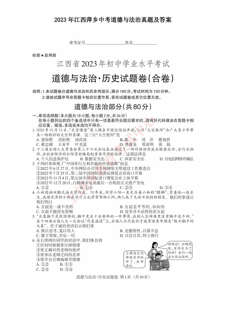 2023年江西萍乡中考道德与法治真题及答案.doc
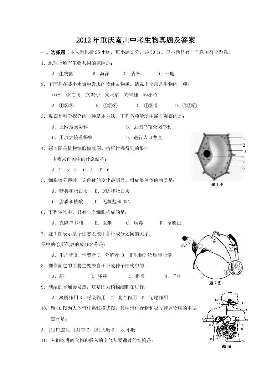 2012年重庆南川中考生物真题及答案.doc
 2013年江西师范大学地理学综合及文艺理论基础考研真题.doc
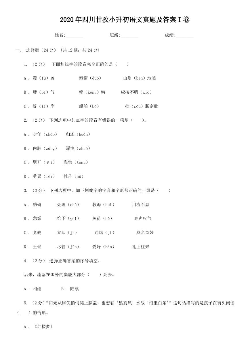 2020年四川甘孜小升初语文真题及答案I卷.doc
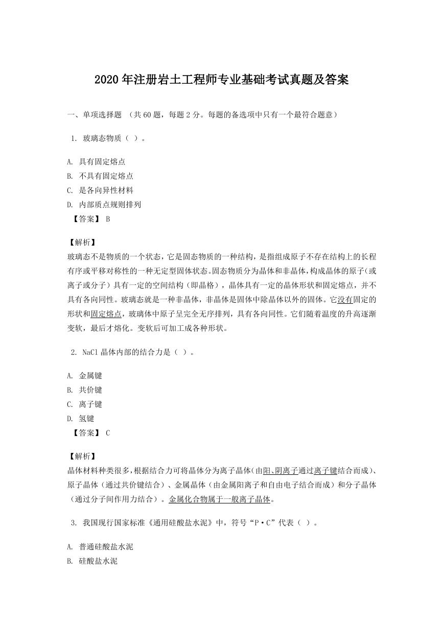 2020年注册岩土工程师专业基础考试真题及答案.doc
 2023-2024学年福建省厦门市九年级上学期数学月考试题及答案.doc
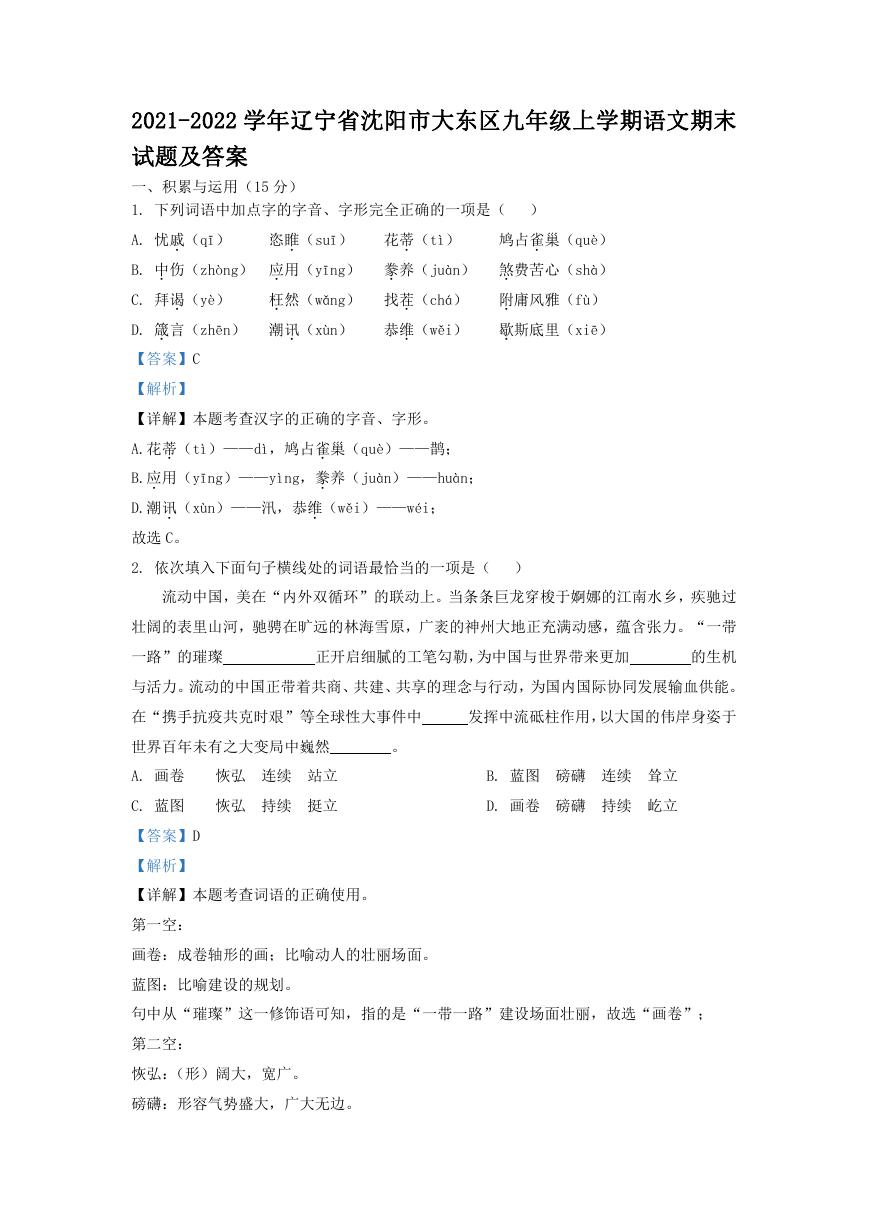 2021-2022学年辽宁省沈阳市大东区九年级上学期语文期末试题及答案.doc
 2022-2023学年北京东城区初三第一学期物理期末试卷及答案.doc
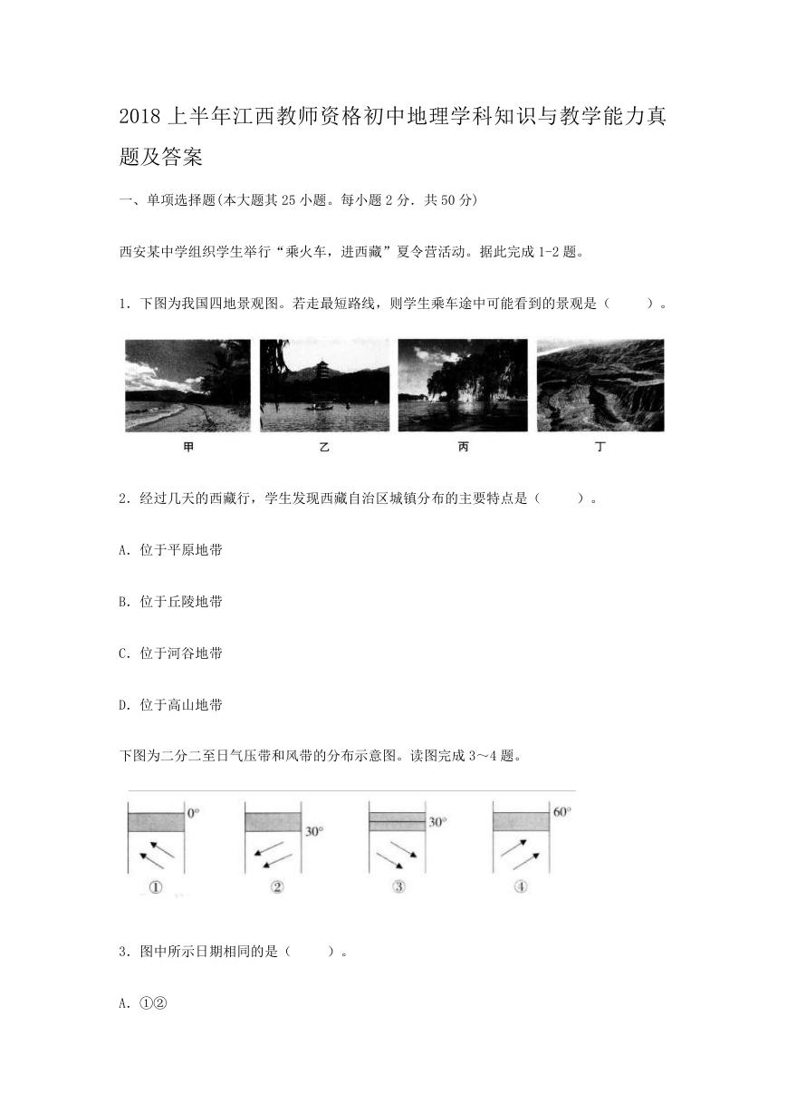 2018上半年江西教师资格初中地理学科知识与教学能力真题及答案.doc
 2012年河北国家公务员申论考试真题及答案-省级.doc
 2020-2021学年江苏省扬州市江都区邵樊片九年级上学期数学第一次质量检测试题及答案.doc
 2022下半年黑龙江教师资格证中学综合素质真题及答案.doc