Preface
Organization
Contents – Part II
Optical and Histology Applications: Optical Imaging Applications
Instance Segmentation and Tracking with Cosine Embeddings and Recurrent Hourglass Networks
1 Introduction
2 Instance Segmentation and Tracking
2.1 Recurrent Stacked Hourglass Network
2.2 Cosine Embedding Loss
2.3 Clustering of Embeddings
3 Experimental Setup and Results
4 Discussion and Conclusion
References
Skin Lesion Classification in Dermoscopy Images Using Synergic Deep Learning
1 Introduction
2 Datasets
3 Method
3.1 Input Layer
3.2 Dual DCNN Components
3.3 Synergic Network
3.4 Training and Testing
4 Results
5 Discussion
6 Conclusion
References
SLSDeep: Skin Lesion Segmentation Based on Dilated Residual and Pyramid Pooling Networks
1 Introduction
2 Proposed Model
2.1 Network Architecture
2.2 Loss Function
3 Experimental Setup and Evaluation
4 Conclusions
References
-Hemolysis Detection on Cultured Blood Agar Plates by Convolutional Neural Networks
1 Introduction
1.1 Problem Definition
1.2 Related Work and Contribution
2 Throat Swab Culture Dataset
3 -Hemolysis Detection Method
3.1 Patch Extraction
3.2 Patch Classification
4 Results and Discussion
5 Conclusion
References
A Pixel-Wise Distance Regression Approach for Joint Retinal Optical Disc and Fovea Detection
1 Introduction
2 Joint Optic Disc and Fovea Detection Methodology
2.1 Casting the Problem into a Pixel-Wise Regression Task
2.2 A Fully-Convolutional Deep Neural Network for Distance Regression
2.3 Optic Disc and the Fovea Assignment
3 Experimental Evaluation
3.1 Data
3.2 Evaluation Approach
3.3 Quantitative Evaluation
4 Conclusions and Future Work
References
Deep Random Walk for Drusen Segmentation from Fundus Images
1 Introduction
2 Deep Random Walk Networks
2.1 Deep Feature Extraction Module
2.2 Affinity Learning Module
2.3 Random Walk Module
3 Implementation
4 Experiment
4.1 Dataset
4.2 Evaluation and Result
5 Conclusion
References
Retinal Artery and Vein Classification via Dominant Sets Clustering-Based Vascular Topology Estimation
1 Introduction
2 Method
2.1 Graph Generation
2.2 Dominant Sets Clustering-Based Topology Estimation
2.3 Artery/Vein Classification
3 Experimental Results
3.1 Topology Estimation
3.2 A/V Classification
4 Conclusions
References
Towards a Glaucoma Risk Index Based on Simulated Hemodynamics from Fundus Images
1 Introduction
2 Materials and Methods
2.1 Preprocessing
2.2 Simulation of the Retinal Hemodynamics
2.3 Bag of Hemodynamic Features (BoHF)
3 Results
4 Discussion
References
A Framework for Identifying Diabetic Retinopathy Based on Anti-noise Detection and Attention-Based Fusion
1 Introduction
2 Method
2.1 Center-Sample Detector
2.2 Attention Fusion Network
3 Experiments
3.1 Evaluating Center-Sample Detector
3.2 Evaluating the Attention Fusion Network
4 Conclusions
References
Deep Supervision with Additional Labels for Retinal Vessel Segmentation Task
1 Introduction
2 Proposed Method
2.1 U-Net
2.2 Additional Label
2.3 Deep Supervision
3 Experiments
3.1 Datasets
3.2 Results
3.3 Comparison
4 Conclusion
References
A Multi-task Network to Detect Junctions in Retinal Vasculature
1 Introduction
2 Method
2.1 Learning Junction Patterns with Multi-task Network
2.2 Initial Junction Search
2.3 Refined Junction Search
3 Experimental Setup and Material
4 Results
5 Conclusion
References
A Multitask Learning Architecture for Simultaneous Segmentation of Bright and Red Lesions in Fundus Images
1 Introduction
2 Methods
2.1 Multitask Architecture
2.2 Training
3 Experiments
4 Results and Discussion
5 Conclusion
References
Uniqueness-Driven Saliency Analysis for Automated Lesion Detection with Applications to Retinal Diseases
1 Introduction
2 Method
3 Experimental Evaluation
3.1 Dark Lesion Detection
3.2 Bright Lesion Detection
4 Conclusions
References
Multiscale Network Followed Network Model for Retinal Vessel Segmentation
1 Introduction
2 Datasets
3 Method
3.1 Images Pre-processing and Patch Extraction
3.2 Training Two NFN Models
3.3 Testing the MS-NFN Model
4 Results
5 Conclusions
References
Optical and Histology Applications: Histology Applications
Predicting Cancer with a Recurrent Visual Attention Model for Histopathology Images
1 Introduction
2 Method
3 Experiments
4 Conclusion
References
A Deep Model with Shape-Preserving Loss for Gland Instance Segmentation
1 Introduction
2 Method
2.1 Shape-Preserving Loss
2.2 Deep Convolutional Neural Network
3 Evaluation and Discussion
4 Conclusion
References
Model-Based Refinement of Nonlinear Registrations in 3D Histology Reconstruction
1 Introduction
2 Methods
2.1 Probabilistic Framework
2.2 Inference: Proposed Method
3 Experiments and Results
3.1 Data
3.2 Experiments on Synthetic Dataset
3.3 Experiments on Allen Dataset
4 Discussion and Conclusion
References
Invasive Cancer Detection Utilizing Compressed Convolutional Neural Network and Transfer Learning
1 Introduction
2 Methodology
2.1 Overview
2.2 Small Capacity Network
2.3 Transfer Learning from Large Capacity Network
2.4 Efficient Inference
3 Experiment and Discussion
4 Conclusion
References
Which Way Round? A Study on the Performance of Stain-Translation for Segmenting Arbitrarily Dyed Histological Images
1 Motivation
2 Methods
2.1 Stain-Translation Model and Sampling Strategies
2.2 Segmentation Model and Evaluation Details
3 Results
4 Discussion
References
Graph CNN for Survival Analysis on Whole Slide Pathological Images
1 Introduction
2 Methodology
3 Experiment
3.1 Dataset
3.2 State-of-the-Art Methods
3.3 Result and Discussion
4 Conclusion
References
Fully Automated Blind Color Deconvolution of Histopathological Images
1 Introduction
2 Problem Formulation
3 Bayesian Modelling and Inference
4 Experiments
5 Conclusions
References
Improving Whole Slide Segmentation Through Visual Context - A Systematic Study
1 Introduction
2 Related Work
3 Methods
4 Results and Discussion
5 Conclusions
References
Adversarial Domain Adaptation for Classification of Prostate Histopathology Whole-Slide Images
1 Introduction
2 Method
3 Experimental Validation and Results
4 Conclusion
References
Rotation Equivariant CNNs for Digital Pathology
1 Introduction
2 Methods
2.1 Background
2.2 G-CNN DenseNet Architecture
3 Experimental Results
3.1 Datasets and Evaluation
3.2 Model Reliability
3.3 P4M-DenseNet Performance
4 Conclusion
References
A Probabilistic Model Combining Deep Learning and Multi-atlas Segmentation for Semi-automated Labelling of Histology
1 Introduction
2 Methods
2.1 Probabilistic Model
2.2 Inference: Proposed Method
2.3 Model Instantiation
3 Experiments and Results
3.1 Data
3.2 Experimental Setup
3.3 Results
4 Discussion and Conclusion
References
BESNet: Boundary-Enhanced Segmentation of Cells in Histopathological Images
1 Introduction
2 Method
2.1 Boundary-Enhanced Segmentation Network (BESNet)
2.2 Boundary-Enhanced Cross-Entropy (BECE) Loss
2.3 Training and Testing
3 Experiments
4 Results and Discussions
5 Conclusions
References
Panoptic Segmentation with an End-to-End Cell R-CNN for Pathology Image Analysis
1 Introduction
2 Methods
2.1 Semantic Segmentation Branch and Feature Map Branch
2.2 Region Proposal Network and Instance Branch
3 Experiment
4 Conclusions
References
Integration of Spatial Distribution in Imaging-Genetics
1 Introduction
2 Method
2.1 Extraction of Image Features
2.2 Computation of Spatial Descriptor
2.3 Canonical Correlation Analysis
3 Experiments and Results
3.1 Ripley's K-Function on Real Data
3.2 CCA with Image and Spatial Features
3.3 Sparse CCA with Image and Spatial Features
4 Discussion and Conclusions
References
Multiple Instance Learning for Heterogeneous Images: Training a CNN for Histopathology
1 Introduction
2 Background
3 Multiple Instance Learning with a CNN
4 Multiple Instance Aggregation
5 Training with Multiple Instance Augmentation
6 Experiments
7 Discussion
References
Optical and Histology Applications: Microscopy Applications
Cell Detection with Star-Convex Polygons
1 Introduction
2 Method
2.1 Implementation
3 Experiments
3.1 Datasets
3.2 Evaluation Metric
3.3 Compared Methods
3.4 Results
4 Discussion
References
Deep Convolutional Gaussian Mixture Model for Stain-Color Normalization of Histopathological Images
1 Introduction
2 Methods
3 Experimental Results
4 Conclusions
References
Learning to Segment 3D Linear Structures Using Only 2D Annotations
1 Introduction
2 Related Work
3 Method
3.1 From 3D to 2D Annotations
3.2 Visual Hull for Training on Cropped Volumes
3.3 Implementation
4 Experimental Evaluation
4.1 Data and Annotations
4.2 User Study
4.3 3D vs 2D Annotations
5 Conclusion
References
A Multiresolution Convolutional Neural Network with Partial Label Training for Annotating Reflectance Confocal Microscopy Images of Skin
1 Introduction
2 Related Work
3 Proposed Model
3.1 Architecture
3.2 Loss Function
4 Dataset and Experiments
References
Weakly-Supervised Learning-Based Feature Localization for Confocal Laser Endomicroscopy Glioma Images
Abstract
1 Introduction
2 Methods
2.1 New Design of Class Activation Map (CAM)
2.2 Lateral Inhibition and Collateral Integration of Localizer Neurons
3 Experimental Setup and Results
4 Conclusions
Acknowledgement
References
Synaptic Partner Prediction from Point Annotations in Insect Brains
1 Introduction
2 Method
2.1 Directed edges for synaptic partner representation
2.2 Edge classification
2.3 Synaptic partner extraction
3 Results
4 Discussion
References
Synaptic Cleft Segmentation in Non-isotropic Volume Electron Microscopy of the Complete Drosophila Brain
1 Introduction
2 Related Work
3 Methods
3.1 Training Setup
3.2 Experiments
3.3 Synaptic Cleft Prediction on the Complete Drosophila Brain
4 Conclusion
References
Weakly Supervised Representation Learning for Endomicroscopy Image Analysis
1 Introduction
2 Methodology
2.1 Frame-Based Feature Representation
2.2 Local Label Classification
2.3 Global Label Classification
2.4 Semantic Exclusivity Loss
2.5 Final Objective and Alternative Learning
3 Experiments
4 Conclusion
References
DeepHCS: Bright-Field to Fluorescence Microscopy Image Conversion Using Deep Learning for Label-Free High-Content Screening
1 Introduction
2 Method
2.1 Data
2.2 Proposed Method: DeepHCS
3 Results
4 Conclusion
References
Optical and Histology Applications: Optical Coherence Tomography and Other Optical Imaging Applications
A Cascaded Refinement GAN for Phase Contrast Microscopy Image Super Resolution
1 Introduction
2 Related Work and Our Proposal
3 Preliminaries
3.1 Generative Adversarial Networks
3.2 Optics-Related Data Enhancement
4 A Cascaded Refinement GAN for Super Resolution
4.1 Network Architecture
4.2 Loss Function
4.3 Implementation and Training Details
5 Experimental Results
6 Conclusion
References
Multi-context Deep Network for Angle-Closure Glaucoma Screening in Anterior Segment OCT
1 Introduction
2 Proposed Method
2.1 AS-OCT Segmentation and Clinical Parameter
2.2 Multi-context Deep Network Architecture
2.3 Data Augmentation for AS-OCT
3 Experiments
4 Conclusion
References
Analysis of Morphological Changes of Lamina Cribrosa Under Acute Intraocular Pressure Change
1 Introduction
2 Methods
2.1 IOP Experiment Setup
2.2 Image Preprocessing
2.3 Unbiased Atlas Building
3 Results
3.1 Validation
3.2 Clinical Study
4 Conclusions
References
Beyond Retinal Layers: A Large Blob Detection for Subretinal Fluid Segmentation in SD-OCT Images
1 Introduction
2 Methodology
2.1 Aggregate Response Construction
2.2 Hessian Analysis
2.3 Post-pruning
3 Results
4 Conclusion
References
Automated Choroidal Neovascularization Detection for Time Series SD-OCT Images
1 Introduction
2 Method
2.1 Method Overview
2.2 Preprocessing
2.3 Classification of positive and negative samples
2.4 3D-HOG Feature Extraction
2.5 Similarity measurement and model update
2.6 Post-processing
3 Experiments
3.1 Quantitative Evaluation
3.2 Qualitative Analysis
4 Conclusions
References
CapsDeMM: Capsule Network for Detection of Munro's Microabscess in Skin Biopsy Images
1 Introduction
2 Proposed Methodology
3 Dataset
4 Experiments
4.1 Experimental Setting
4.2 Results and Discussion
5 Conclusion and Future Work
References
Webly Supervised Learning for Skin Lesion Classification
1 Introduction
2 Methodology
2.1 Model Learning
3 Experiments
4 Results and Discussion
5 Conclusions
References
Feature Driven Local Cell Graph (FeDeG): Predicting Overall Survival in Early Stage Lung Cancer
Abstract
1 Introduction
2 Brief Overview and Novel Contributions
3 Feature Driven Cell Graph
3.1 Nuclei Segmentation and Morphologic Feature Extraction
3.2 FeDeG Construction in Nuclear Morphologic Feature Space
3.3 FeDeG Features Computation
4 Experimental Design
4.1 Dataset Description
4.2 Comparative Methods
4.2.1 Graph Based and Other Pathomic Strategies
4.2.2 Deep Learning
5 Results and Discussion
5.1 Discrimination of Different Graph and Deep Learning Representations
5.2 Survival Analysis
6 Concluding Remarks
References
Cardiac, Chest and Abdominal Applications: Cardiac Imaging Applications
Towards Accurate and Complete Registration of Coronary Arteries in CTA Images
1 Introduction
2 Methods
2.1 Bifurcation Matching
2.2 Segment Registration
2.3 Further Segmentation and Registration
3 Experiments and Results
4 Discussion and Conclusion
References
Quantifying Tensor Field Similarity with Global Distributions and Optimal Transport
1 Introduction
2 Background
3 Quantifying Tensor Field Similarity
4 Experiments and Results
5 Discussion
References
Cardiac Motion Scoring with Segment- and Subject-Level Non-local Modeling
1 Introduction
2 Cardiac Motion Scoring
2.1 Myocardium Segment Re-sampling in Polar Coordinate System
2.2 Motion Scoring Neural Network
3 Dataset and Configurations
4 Results and Analysis
5 Conclusions
References
Computational Heart Modeling for Evaluating Efficacy of MRI Techniques in Predicting Appropriate ICD Therapy
Abstract
1 Introduction
2 Methods
2.1 Study Subjects and Image Acquisition
2.2 Image Processing
2.3 Simulation of Cardiac Electrophysiology
3 Results
4 Discussion and Conclusion
References
Multiview Two-Task Recursive Attention Model for Left Atrium and Atrial Scars Segmentation
1 Introduction
2 Method
3 Experimental Results and Discussion
4 Conclusion
References
Learning Interpretable Anatomical Features Through Deep Generative Models: Application to Cardiac Remodeling
1 Introduction
1.1 Related Work
2 Materials and Methods
2.1 Datasets
2.2 Deep Generative Model
3 Results
4 Discussion and Conclusion
References
Joint Learning of Motion Estimation and Segmentation for Cardiac MR Image Sequences
1 Introduction
1.1 Related Work
2 Methods
2.1 Unsupervised Cardiac Motion Estimation
2.2 Joint Model for Cardiac Motion Estimation and Segmentation
3 Experiments and Results
4 Conclusion
References
Multi-Input and Dataset-Invariant Adversarial Learning (MDAL) for Left and Right-Ventricular Coverage Estimation in Cardiac MRI
1 Introduction
2 Methodology
2.1 Problem Formulation
2.2 Multi-Input and Dataset-Invariant Adversarial Learning
2.3 Optimization
2.4 Detection and Regression for Basal/Apical Slice Position
3 Experiments and Analysis
4 Conclusion
References
Factorised Spatial Representation Learning: Application in Semi-supervised Myocardial Segmentation
1 Introduction
2 Related Work
3 Proposed Approach: The SDNet
4 Experiments and Discussion
4.1 Data and Baselines
4.2 Latent Space Arithmetic
4.3 Semi-supervised Results
5 Conclusion
References
High-Dimensional Bayesian Optimization of Personalized Cardiac Model Parameters via an Embedded Generative Model
1 Introduction
2 Background: Cardiac Electrophysiological System
3 HD Parameter Estimation
3.1 LD-to-HD Parameter Generation via VAE
3.2 Bayesian Optimization with Embedded Generative Model
4 Experiments
References
Generative Modeling and Inverse Imaging of Cardiac Transmembrane Potential
1 Introduction
2 Generative Modeling of TMP via Sequential VAE
3 Transmural EP Imaging
4 Results
5 Discussion and Conclusions:
References
Pulmonary Vessel Tree Matching for Quantifying Changes in Vascular Morphology
1 Introduction
2 Methods
2.1 Vascular Tree Construction
2.2 Vascular Tree Matching
2.3 Quantitative Analysis
3 Experiment
4 Results
5 Discussion and Conclusion
References
MuTGAN: Simultaneous Segmentation and Quantification of Myocardial Infarction Without Contrast Agents via Joint Adversarial Learning
1 Introduction
2 Methodology
2.1 MuTGAN Formulation
2.2 Generator
2.3 Discriminator
3 Materials and Implementation Details
4 Experiments and Results
5 Conclusions
References
More Knowledge Is Better: Cross-Modality Volume Completion and 3D+2D Segmentation for Intracardiac Echocardiography Contouring
1 Introduction
2 Method
2.1 3D Sparse Volume Segmentation and Completion
2.2 2D Contour Refinement
3 Experiments
4 Conclusions and Future Work
References
Unsupervised Domain Adaptation for Automatic Estimation of Cardiothoracic Ratio
1 Introduction
2 Methodology
2.1 Adversarial Training for Supervised Semantic Segmentation
2.2 Unsupervised Domain Adaption
2.3 Estimation of CTR
2.4 Semi-Supervised Semantic Segmentation
3 Experimental Results
4 Conclusions
References
TextRay: Mining Clinical Reports to Gain a Broad Understanding of Chest X-Rays
1 Introduction
2 Materials and Methods
2.1 Model
2.2 Evaluation Sets
3 Results
4 Discussion
5 Conclusion
References
Localization and Labeling of Posterior Ribs in Chest Radiographs Using a CRF-regularized FCN with Local Refinement
1 Introduction
2 Method
2.1 Generating Localization Hypotheses Using a U-Net
2.2 Selecting Reasonable Localization Hypotheses Using a CRF
2.3 Going Beyond Potentially Incorrect Localization Hypotheses
3 Experiments and Results
4 Discussion and Conclusions
References
Evaluation of Collimation Prediction Based on Depth Images and Automated Landmark Detection for Routine Clinical Chest X-Ray Exams
Abstract
1 Introduction
2 Methods
2.1 Data Pre-processing
2.2 Annotation of Reference Lung Landmarks and Lung Bounding Box
2.3 Trained Classifier for the Detection of Anatomical Landmarks
2.4 Statistical Models for the Computation of Collimation Parameters
3 Results
4 Discussion/Conclusions
References
Efficient Active Learning for Image Classification and Segmentation Using a Sample Selection and Conditional Generative Adversarial Network
1 Introduction
2 Methods
2.1 Conditional Generative Adversarial Networks
2.2 Sample Informativeness Using Uncertainty Form Bayesian Neural Networks
2.3 Implementation Details
3 Experiments
3.1 Classification Results
3.2 Segmentation Performance
3.3 Savings in Annotation Effort
4 Conclusion
References
Iterative Attention Mining for Weakly Supervised Thoracic Disease Pattern Localization in Chest X-Rays
1 Introduction
2 Methods
2.1 Disease Pattern Localization with Attention Mining
2.2 Incremental Learning with Knowledge Preservation
2.3 Multi-scale Aggregation
3 Experimental Results and Analysis
3.1 Multiple Scale Aggregation
3.2 Disease Pattern Localization with AM and KP
4 Conclusion
References
Task Driven Generative Modeling for Unsupervised Domain Adaptation: Application to X-ray Image Segmentation
1 Introduction
2 Methodology
2.1 Problem Overview
2.2 Dense Image to Image Network for Segmentation on DRRs
2.3 Task Driven Generative Adversarial Networks (TD-GAN)
3 Experiments and Results
4 Discussions and Conclusions
References
Cardiac, Chest and Abdominal Applications: Colorectal, Kidney and Liver Imaging Applications
Towards Automated Colonoscopy Diagnosis: Binary Polyp Size Estimation via Unsupervised Depth Learning
1 Introduction
2 Methods
2.1 Spatio-temporal Video Based Polyp Detection
2.2 Two-Category Polyp Size Estimation
3 Experimental Results
3.1 Polyp Detection
3.2 Polyp Size Estimation
4 Discussion
5 Conclusions
References
RIIS-DenseNet: Rotation-Invariant and Image Similarity Constrained Densely Connected Convolutional Network for Polyp Detection
1 Introduction
2 Method: RIIS-DenseNet Model
2.1 Data Rotation Augmentation
2.2 DenseNet
2.3 Joint Loss Function
3 Experiment Setup and Results
4 Conclusion
References
Interaction Techniques for Immersive CT Colonography: A Professional Assessment
1 Introduction
2 Immersive CT Colonography System
2.1 Apparatus
2.2 3D Data
2.3 Interaction Design
3 Evaluation with Professionals
4 Conclusions
References
Quasi-automatic Colon Segmentation on T2-MRI Images with Low User Effort
1 Introduction
2 Method Overview
2.1 Tubularity Detection Filter
3 Evaluation and Results
4 Conclusions
References
Ordinal Multi-modal Feature Selection for Survival Analysis of Early-Stage Renal Cancer
1 Introduction
2 Method
3 Experimental Results
4 Conclusion
References
Noninvasive Determination of Gene Mutations in Clear Cell Renal Cell Carcinoma Using Multiple Instance Decisions Aggregated CNN
1 Introduction
2 Materials and Methods
2.1 Data
2.2 Multiple Instance Decision Aggregation for Mutation Detection
3 Results
4 Conclusions
References
Combining Convolutional and Recurrent Neural Networks for Classification of Focal Liver Lesions in Multi-phase CT Images
Abstract
1 Introduction
2 Methodology
2.1 ResGLNet
2.2 BD-LSTM
2.3 Combining ResGLNet and BD-LSTM
2.4 Training Strategy
2.5 Post-processing of Label Map and Classification of Lesions
3 Experiments
3.1 Data and Implementation
3.2 Results
4 Conclusions
Acknowledgements
Acknowledgements
References
Construction of a Spatiotemporal Statistical Shape Model of Pediatric Liver from Cross-Sectional Data
1 Introduction
2 Methods
2.1 PCA with Temporal Regularization
2.2 Data Augmentation
3 Experiments
4 Conclusion
References
Deep 3D Dose Analysis for Prediction of Outcomes After Liver Stereotactic Body Radiation Therapy
1 Introduction
2 Methodology
2.1 Registration of 3D Dose Plans into Anatomy-Unified Space
2.2 CNN Transfer Learning for Outcome Prediction After Liver SBRTs
2.3 Treatment Outcome Atlases
3 Experiments and Results
3.1 Post-SBRT Survival and Local Progression Prediction
3.2 Survival and Local Progression Atlases
4 Discussion and Conclusion
References
Liver Lesion Detection from Weakly-Labeled Multi-phase CT Volumes with a Grouped Single Shot MultiBox Detector
1 Introduction
2 Multi-phase Data
3 Grouped Single Shot MultiBox Detector
4 Experiments
5 Discussion and Conclusions
References
A Diagnostic Report Generator from CT Volumes on Liver Tumor with Semi-supervised Attention Mechanism
1 Introduction
2 Model
3 Experiments
4 Conclusion
References
Less is More: Simultaneous View Classification and Landmark Detection for Abdominal Ultrasound Images
1 Introduction
2 Methods and Results
2.1 Data and Task Definitions
2.2 MTL Framework
2.3 Results
3 Discussion
References
Cardiac, Chest and Abdominal Applications: Lung Imaging Applications
Deep Active Self-paced Learning for Accurate Pulmonary Nodule Segmentation
1 Introduction
2 Method
2.1 Nodule R-CNN
2.2 The Deep Active Self-paced Learning Strategy
3 Experiments and Results
4 Conclusions
References
CT-Realistic Lung Nodule Simulation from 3D Conditional Generative Adversarial Networks for Robust Lung Segmentation
1 Introduction
2 Methods
2.1 CGAN Formulation
2.2 3D CGAN Architecture
2.3 CGAN Optimization
3 Experiments and Results
3.1 3D CGAN Performance
3.2 Improving Pathological Lung Segmentation
4 Conclusion
References
Fast CapsNet for Lung Cancer Screening
1 Introduction
2 Capsule Network
3 Fast Capsule Network
4 Experiments
5 Conclusions
References
Mean Field Network Based Graph Refinement with Application to Airway Tree Extraction
1 Introduction
2 Method
2.1 The Graph Refinement Model
2.2 Mean Field Network
2.3 Airway Tree Extraction as Graph Refinement
3 Experiments and Results
4 Discussion and Conclusion
References
Automated Pulmonary Nodule Detection: High Sensitivity with Few Candidates
1 Introduction
2 Methods
2.1 High Sensitivity Candidate Detection with FPN
2.2 Conditional 3D-NMS for Redundant Candidate Removal
2.3 Attention 3D-CNN for False Positive Reduction
3 Experiments
3.1 Implementation Details
3.2 Ablation Study and Results
4 Conclusion
References
Deep Learning from Label Proportions for Emphysema Quantification
1 Introduction
2 Methods
2.1 Architectures
2.2 A Loss for Learning from Proportion Intervals (LPI)
3 Experimental Setting
4 Results
5 Discussion and Conclusion
References
Tumor-Aware, Adversarial Domain Adaptation from CT to MRI for Lung Cancer Segmentation
1 Introduction
2 Method
2.1 Step 1: MRI Synthesis Using Tumor-Aware Unsupervised Cross Domain Adaptation
2.2 Step 2: Semi-supervised Tumor Segmentation from MRI
2.3 Network Structure and Implementation
3 Experiments and Results
3.1 Ablation Tests
3.2 Datasets
3.3 MR Image Synthesis Results
3.4 Segmentation Results
4 Discussion
5 Conclusions
References
From Local to Global: A Holistic Lung Graph Model
1 Introduction
2 Methods
2.1 Dataset
2.2 Holistic Graph Model of the Lungs
2.3 Undirected Weighted Graph Model of the Lung
2.4 Directed Weighted Graph-Model of the Lungs
2.5 Graph-Based Patient Descriptor
3 Experimental Setup
4 Results
5 Discussion
6 Conclusions and Future Work
References
S4ND: Single-Shot Single-Scale Lung Nodule Detection
1 Introduction
2 Method
2.1 Single-Scale Detection
2.2 Dense and Deeper Convolution Blocks Improve Detection
2.3 Max-Pooling Improves Detection
2.4 Proposed 3D Deep Network Architecture
3 Experiments and Results
4 Conclusion
References
Vascular Network Organization via Hough Transform (VaNgOGH): A Novel Radiomic Biomarker for Diagnosis and Treatment Response
1 Introduction
2 Previous Work and Novel Contributions
3 Methodology
3.1 Notation
3.2 VaNgOGH Descriptor
4 Experimental Results and Discussion
4.1 Data Description
4.2 Comparative Strategies and Classifier Construction
4.3 Experiment 1: Pre-treatment Response Prediction in Breast Cancer DCE-MRI
4.4 Experiment 2: Malignancy Diagnosis for Lung Nodules
5 Concluding Remarks
References
DeepEM: Deep 3D ConvNets with EM for Weakly Supervised Pulmonary Nodule Detection
1 Introduction
2 DeepEM for Weakly Supervised Detection
3 Experiments
4 Conclusion
References
Statistical Framework for the Definition of Emphysema in CT Scans: Beyond Density Mask
1 Introduction
2 Characterization of Emphysema in CT Scans
3 Adaptive Threshold for Emphysema Detection
4 Results
5 Conclusion
References
Cardiac, Chest and Abdominal Applications: Breast Imaging Applications
Conditional Generative Adversarial and Convolutional Networks for X-ray Breast Mass Segmentation and Shape Classification
1 Introduction
2 Related Work
3 Proposed Model
3.1 System Overview
3.2 Mass Segmentation Model (with cGAN)
3.3 Shape classification model (with CNN)
4 Experiments
4.1 Datasets
4.2 Experimental Results
5 Conclusions
References
A Robust and Effective Approach Towards Accurate Metastasis Detection and pN-stage Classification in Breast Cancer
1 Introduction
2 Methodology
2.1 Regions of Interests Extraction
2.2 Metastasis Detection
2.3 Lymph Node Classification
3 Experiments
3.1 Dataset
3.2 Evaluation Metrics
3.3 Experimental Details
3.4 Results
4 Conclusion
References
3D Anisotropic Hybrid Network: Transferring Convolutional Features from 2D Images to 3D Anisotropic Volumes
1 Introduction
2 Anisotropic Hybrid Network
2.1 Pre-training a Multi-Channel 2D Feature Encoder
2.2 Transferring the Learned 2D Features into 3D AH-Net
2.3 Anisotropic Hybrid Decoder
2.4 Training the AH-Net
3 Experimental Results
3.1 Breast Lesion Detection from DBT
3.2 Liver and Liver Tumor Segmentation from CT
4 Conclusion
References
Deep Generative Breast Cancer Screening and Diagnosis
1 Introduction
2 Methods
2.1 Model Overview
2.2 GAN-Enhanced Deep Classification
2.3 Training
2.4 Transfer Learning
3 Experiments and Results
3.1 Performance Enhanced by GAN
4 Conclusion
References
Integrate Domain Knowledge in Training CNN for Ultrasonography Breast Cancer Diagnosis
Abstract
1 Introduction
2 Method
2.1 Data Acquisition
2.2 Image Preprocessing
2.3 Training Multi-task CNN
3 Results
3.1 Classification Result
3.2 Examples of Correct and Wrong Predictions
4 Conclusion
Acknowledgement
References
Small Lesion Classification in Dynamic Contrast Enhancement MRI for Breast Cancer Early Detection
1 Introduction
2 Methods
2.1 Datasets
2.2 DC-LSTM
2.3 Latent Attributes Learning
3 Experiments
3.1 DC-LSTM
3.2 Latent Attributes Learning
4 Conclusion
References
Thermographic Computational Analyses of a 3D Model of a Scanned Breast
1 Introduction
2 Materials and Methods
2.1 Heat Biotransference Equation
2.2 3D Scanned Computational Model and Numerical Solution
2.3 Case of Simulated Breast Cancer
3 Results and Discussions
4 Conclusions
References
Y-Net: Joint Segmentation and Classification for Diagnosis of Breast Biopsy Images
1 Introduction
2 A System for Joint Segmentation and Classification
2.1 Y-Net Architecture
2.2 Discriminative Instance Selection
2.3 Diagnostic Classification
3 Experiments
3.1 Segmentation Results
3.2 Diagnostic Classification Results
4 Conclusion
References
Cardiac, Chest and Abdominal Applications: Other Abdominal Applications
AutoDVT: Joint Real-Time Classification for Vein Compressibility Analysis in Deep Vein Thrombosis Ultrasound Diagnostics
1 Introduction
2 Method
3 Data and Experiments
4 Results and Discussion
References
MRI Measurement of Placental Perfusion and Fetal Blood Oxygen Saturation in Normal Pregnancy and Placental Insufficiency
1 Introduction
2 Methods
3 Results
4 Discussion
References
Automatic Lacunae Localization in Placental Ultrasound Images via Layer Aggregation
1 Introduction
2 Methods
2.1 From Dot Annotation to Confidence Map
2.2 Lacunae Localization: Layer Aggregation Approaches
3 Experiments
4 Discussion
References
A Decomposable Model for the Detection of Prostate Cancer in Multi-parametric MRI
1 Introduction
2 Related Works
3 Methods
3.1 Random Ferns
3.2 Random Fern Decomposition
3.3 Data
3.4 Prostate Segmentation
3.5 Training
3.6 Statistical Analysis
4 Results
5 Discussion
6 Conclusion
References
Direct Automated Quantitative Measurement of Spine via Cascade Amplifier Regression Network
1 Introduction
2 Cascade Amplifier Regression Network
2.1 Mathematical Formulation
2.2 CARN Architecture
2.3 Local Shape-Constrained Manifold Regularization Loss Function
3 Experimental Results
4 Conclusions
References
Estimating Achilles Tendon Healing Progress with Convolutional Neural Networks
1 Introduction
2 Method
3 Experiments
3.1 Dataset
3.2 Tendon Healing Process Assessment
4 Conclusions
References
Author Index




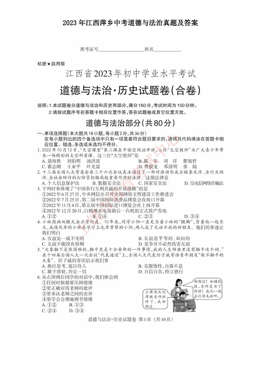 2023年江西萍乡中考道德与法治真题及答案.doc
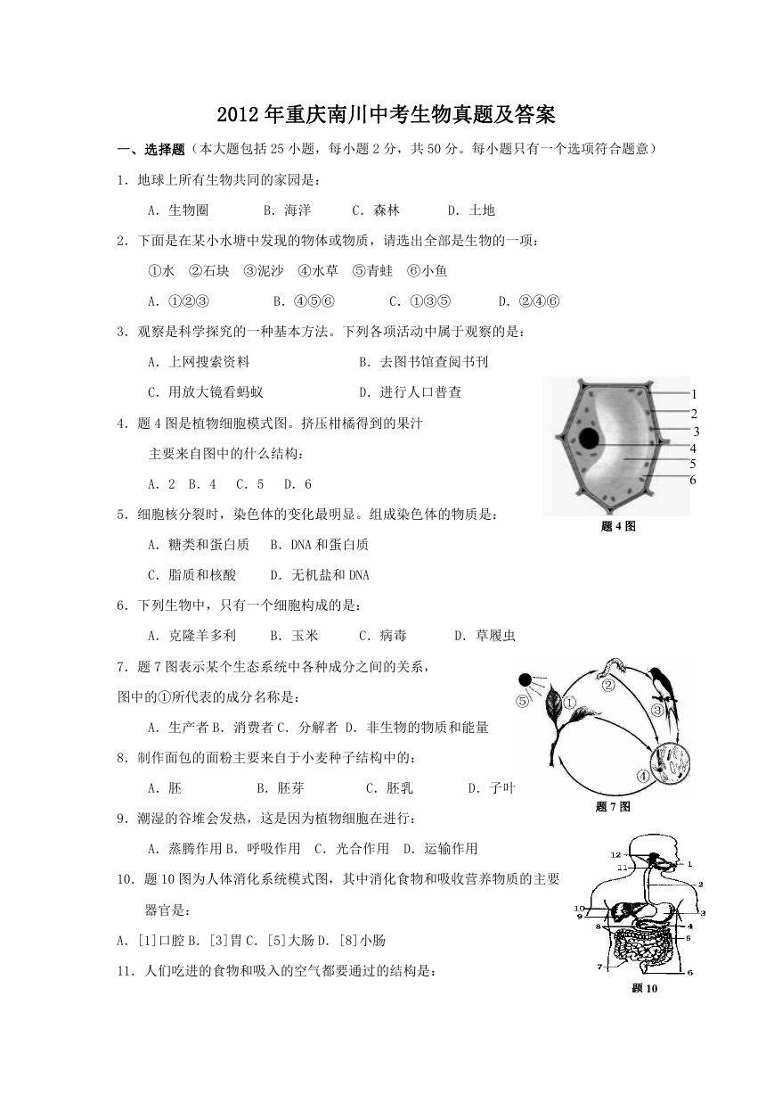 2012年重庆南川中考生物真题及答案.doc
 2013年江西师范大学地理学综合及文艺理论基础考研真题.doc
 2020年四川甘孜小升初语文真题及答案I卷.doc
 2020年注册岩土工程师专业基础考试真题及答案.doc
 2023-2024学年福建省厦门市九年级上学期数学月考试题及答案.doc
 2021-2022学年辽宁省沈阳市大东区九年级上学期语文期末试题及答案.doc
 2022-2023学年北京东城区初三第一学期物理期末试卷及答案.doc
 2018上半年江西教师资格初中地理学科知识与教学能力真题及答案.doc
 2012年河北国家公务员申论考试真题及答案-省级.doc
 2020-2021学年江苏省扬州市江都区邵樊片九年级上学期数学第一次质量检测试题及答案.doc
 2022下半年黑龙江教师资格证中学综合素质真题及答案.doc