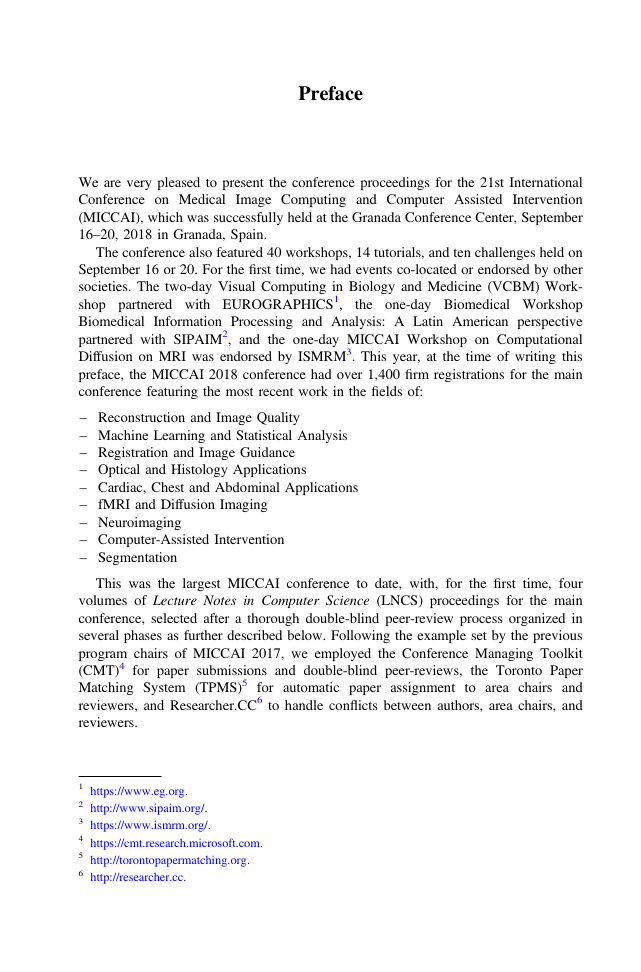
Preface
Organization
Contents – Part IV
Computer Assisted Interventions: Image Guided Interventions and Surgery
Uncertainty in Multitask Learning: Joint Representations for Probabilistic MR-only Radiotherapy Planning
1 Introduction
2 Methods
3 Experiments and Results
4 Conclusions
References
A Combined Simulation and Machine Learning Approach for Image-Based Force Classification During Robotized Intravitreal Injections
1 Introduction
2 Method
2.1 Numerical Simulation for Fast Data Generation
2.2 Neural Network Image Classification
3 Results
3.1 Experimental Set-Up
3.2 Data Generation for Neural Network Training
3.3 Tests on Unseen Data
4 Conclusion and Discussion
References
Learning from Noisy Label Statistics: Detecting High Grade Prostate Cancer in Ultrasound Guided Biopsy
1 Introduction
2 Materials
3 Method
3.1 Discriminative Model
3.2 Cancer Grading and Tumor in Core Length Estimation
3.3 Model Uncertainty Estimation
4 Experiments and Results
5 Conclusion
References
A Feature-Driven Active Framework for Ultrasound-Based Brain Shift Compensation
1 Introduction
2 Method
2.1 The Role of US-to-US Registration
2.2 Feature-Based Registration Strategy
2.3 GP Kernel Estimation
3 Experiments
4 Discussion
References
Soft-Body Registration of Pre-operative 3D Models to Intra-operative RGBD Partial Body Scans
1 Introduction, Background and Contributions
2 Methods
2.1 Biomechanical Model Description
2.2 Intra-operative Patient Scanning and Segmentation
2.3 Registration Problem Formulation
2.4 Optimization and Initialization
3 Experimental Results
3.1 Quantitative Analysis with Porcine Datasets
3.2 Qualitative Analysis with a Human Patient
4 Conclusion
References
Automatic Classification of Cochlear Implant Electrode Cavity Positioning
Abstract
1 Introduction
2 Methods
3 Results
4 Conclusions
Acknowledgements
References
X-ray-transform Invariant Anatomical Landmark Detection for Pelvic Trauma Surgery
1 Introduction
2 Method
3 Experiments and Results
3.1 Synthetic X-Rays
3.2 Real X-Rays
4 Discussion and Conclusions
References
Endoscopic Navigation in the Absence of CT Imaging
1 Introduction
2 Method
3 Experimental Results and Discussion
3.1 Experiment 1: Simulation
3.2 Experiment 2: In-Vivo
4 Conclusion
References
A Novel Mixed Reality Navigation System for Laparoscopy Surgery
Abstract
1 Introduction
2 Mixed Reality Navigation for Laparoscopy Surgery
2.1 Mixed-Reality Head Mounted Display (HMD)
2.2 Mixed Reality Navigation Software
2.3 Audio Navigation System
3 Experimental Methods
4 Results and Discussion
5 Conclusion
References
Respiratory Motion Modelling Using cGANs
1 Introduction
2 Method
2.1 Data Acquisition and Image Registration
2.2 Training of the Neural Network
2.3 Real-Time Prediction of Deformation Fields and Stacking
3 Experiments and Results
4 Discussion and Conclusion
References
Physics-Based Simulation to Enable Ultrasound Monitoring of HIFU Ablation: An MRI Validation
1 Introduction
2 Methods
2.1 Biophysical Modeling of HIFU Thermal Ablation
2.2 Ultrasound Thermal Monitoring
3 Experiments and Results
3.1 Sensitivity Analysis
3.2 Phantom Feasibility Study
4 Discussion and Conclusion
References
DeepDRR – A Catalyst for Machine Learning in Fluoroscopy-Guided Procedures
1 Introduction
2 Methods
2.1 Background and Requirements
2.2 DeepDRR
3 Experiments and Results
3.1 Framework Validation
3.2 Task-Based Evaluation
4 Discussion and Conclusion
References
Exploiting Partial Structural Symmetry for Patient-Specific Image Augmentation in Trauma Interventions
1 Introduction
2 Materials and Methods
2.1 Problem Formulation
2.2 Robust Loss for Estimation of Partial Symmetry
2.3 Bone Density Histogram Regularization
2.4 Patient-Specific Image Augmentation
3 Experimental Validation and Results
4 Discussion and Conclusion
References
Intraoperative Brain Shift Compensation Using a Hybrid Mixture Model
1 Introduction
2 Materials and Methods
3 Experiments and Results
4 Discussion and Conclusion
References
Video-Based Computer Aided Arthroscopy for Patient Specific Reconstruction of the Anterior Cruciate Ligament
1 Introduction
2 Video-Based Computer-Aided Arthroscopy
3 Surgical Workflow and Algorithmic Modules
4 Experiments
4.1 Lens Rotation
4.2 3D Registration
5 Conclusions
References
Simultaneous Segmentation and Classification of Bone Surfaces from Ultrasound Using a Multi-feature Guided CNN
1 Introduction
2 Proposed Method
2.1 Enhancement of Bone Surface and Bone Shadow Information
2.2 Pre-enhancing Network (PE)
2.3 Joint Learning of Classification and Segmentation
2.4 Data Acquisition and Training
3 Experimental Results
4 Conclusion
References
Endoscopic Laser Surface Scanner for Minimally Invasive Abdominal Surgeries
1 Introduction
2 Methods and Materials
2.1 Laser Beam Calibration
2.2 Surface Reconstruction as Line-to-Plane Intersection
2.3 Validation
3 Results
4 Discussion and Conclusion
References
Deep Adversarial Context-Aware Landmark Detection for Ultrasound Imaging
1 Introduction
2 Methods
2.1 Baseline Approach for Landmark Detection
2.2 Multitask Learning for Joint Landmark and Contour Detection
3 Results and Discussion
References
Towards a Fast and Safe LED-Based Photoacoustic Imaging Using Deep Convolutional Neural Network
1 Introduction
2 Methods
2.1 Architecture
2.2 Loss Function
3 Experiments and Materials
3.1 LED Excitation Source
3.2 Data Acquisition
3.3 Materials
3.4 Training
4 Evaluation and Results
4.1 Peak Signal-to-Noise-Ratio and Structural Similarity Index
4.2 Performance at Different Depths
4.3 Qualitative Analysis
4.4 Computation Time
5 Discussion and Conclusion
References
An Open Framework Enabling Electromagnetic Tracking in Image-Guided Interventions
1 Introduction
2 Framework Design
3 Framework Implementation
4 Experiments and Results
4.1 Performance Benchmark
4.2 Accuracy Benchmark
5 Discussion
6 Conclusion
References
Colon Shape Estimation Method for Colonoscope Tracking Using Recurrent Neural Networks
1 Introduction
2 Colon Shape Estimation Method
2.1 Overview
2.2 Colon and Colonoscope Shape Representation
2.3 Colonoscope Shape Features
2.4 Shape Estimation Network
3 Experimental Setup
3.1 Colonoscope Shape Measurement
3.2 Colon Shape Measurement
3.3 Shape Estimation Network Training
3.4 Colon Shape Estimation
3.5 Evaluation Metric
4 Experimental Results
5 Discussion
References
Towards Automatic Report Generation in Spine Radiology Using Weakly Supervised Framework
1 Introduction
2 The Proposed Framework for Report Generation
2.1 Recurrent Generative Adversarial Network
2.2 Prior Knowledge-Based Symbolic Program Synthesis
3 Results
4 Discussion and Conclusion
References
Computer Assisted Interventions: Surgical Planning, Simulation and Work Flow Analysis
A Natural Language Interface for Dissemination of Reproducible Biomedical Data Science
1 Introduction
2 Archetypal Analysis Scenarios
3 The Core System Architecture
4 Conclusion
References
Spatiotemporal Manifold Prediction Model for Anterior Vertebral Body Growth Modulation Surgery in Idiopathic Scoliosis
1 Introduction
2 Method
2.1 Discriminant Embedding of Longitudinal Spine Models
2.2 Piecewise-Geodesic Spatiotemporal Manifold
2.3 Prediction of Spine Correction
3 Experiments
4 Conclusion
References
Evaluating Surgical Skills from Kinematic Data Using Convolutional Neural Networks
1 Introduction
2 Method
2.1 Dataset
2.2 Architecture
2.3 Training and Testing
2.4 Class Activation Map
3 Results
3.1 Surgical Skill Classification
3.2 Feedback Visualization
4 Conclusion
References
Needle Tip Force Estimation Using an OCT Fiber and a Fused convGRU-CNN Architecture
1 Introduction
2 Materials and Methods
2.1 Needle Design and Experimental Setup
2.2 Model Architecture
2.3 Data Acquisition and Datasets
3 Results
4 Discussion
5 Conclusion
References
Fast GPU Computation of 3D Isothermal Volumes in the Vicinity of Major Blood Vessels for Multiprobe Cryoablation Simulation
1 Introduction
2 Materials and Methods
2.1 General Formulation
2.2 Propagation of Cold in the Human Body Near Heating Sources
2.3 Validation in Silico
2.4 Validation on Intraoperative MRI Images
3 Results
4 Conclusion
References
A Machine Learning Approach to Predict Instrument Bending in Stereotactic Neurosurgery
1 Introduction
2 Methods
3 Experimental Design and Validation
4 Conclusion
References
Deep Reinforcement Learning for Surgical Gesture Segmentation and Classification
1 Introduction
2 Preliminary
3 Proposed Method
4 Experiments
5 Conclusion and Future Work
References
Automated Performance Assessment in Transoesophageal Echocardiography with Convolutional Neural Networks
1 Introduction
2 Methods
2.1 Dataset Generation
2.2 CNN Architectures
3 Experimentation and Results
4 Conclusions
References
DeepPhase: Surgical Phase Recognition in CATARACTS Videos
1 Introduction
2 Materials and Methods
2.1 Augmented CATARACT Dataset
2.2 Tool Recognition with CNNs
2.3 Phase Recognition with RNNs
3 Experimental Results
3.1 Evaluation Metrics
3.2 Tool Recognition
3.3 Phase Recognition
4 Discussion and Conclusion
References
Surgical Activity Recognition in Robot-Assisted Radical Prostatectomy Using Deep Learning
1 Introduction
2 Methodology
3 Experimental Evaluation
4 Results and Discussion
5 Conclusion
References
Unsupervised Learning for Surgical Motion by Learning to Predict the Future
1 Introduction
2 Methods
2.1 Recurrent Neural Networks and Long Short-Term Memory
2.2 The RNN Encoder-Decoder
2.3 Mixture Density Networks
2.4 Training
3 Experiments
3.1 Dataset
3.2 Future Prediction
3.3 Information Retrieval with Motion-Based Queries
4 Summary and Future Work
References
Computer Assisted Interventions: Visualization and Augmented Reality
Volumetric Clipping Surface: Un-occluded Visualization of Structures Preserving Depth Cues into Surrounding Organs
1 Introduction
1.1 Focus+Context
1.2 Clinical Application
2 Existing Techniques
2.1 Clipping Planes
2.2 Cut-Aways
3 A Real-Time Depth Contiguous Clipping Surface
3.1 Methodology
3.2 Computational Complexity
4 Simulation
5 Conclusions
References
Closing the Calibration Loop: An Inside-Out-Tracking Paradigm for Augmented Reality in Orthopedic Surgery
1 Introduction
2 Materials and Methods
2.1 Calibration
2.2 Prototype
2.3 Virtual Content in the AR Environment
2.4 Experiments and Feasibility Study
3 Results
4 Discussion and Conclusion
References
Higher Order of Motion Magnification for Vessel Localisation in Surgical Video
1 Introduction
2 Methods
2.1 Temporal Filtering
2.2 Phase-Based Magnification
3 Results
4 Conclusion
References
Simultaneous Surgical Visibility Assessment, Restoration, and Augmented Stereo Surface Reconstruction for Robotic Prostatectomy
1 Endoscopic Vision
2 Approaches
2.1 Visibility Assessment
2.2 Visualization Restoration
2.3 Augmented Disparity
3 Validation
4 Results and Discussion
5 Conclusions
References
Real-Time Augmented Reality for Ear Surgery
1 Introduction
2 Methodology
2.1 Initial Semi-automatic Endosocope-CT Registration
2.2 Endoscope-Target Motion Tracking
2.3 3D Pose Estimation of Surgical Instrument
3 Experimental Setup and Results
4 Conclusion
References
Framework for Fusion of Data- and Model-Based Approaches for Ultrasound Simulation
1 Introduction
2 Methods
2.1 Data-Based: Realistic Background from Images
2.2 Model-Based Simulation: Detailed, Arbitrary Content
2.3 Tissue Deformations
2.4 Texture Filling
2.5 Compounding Contents
3 Results
4 Conclusions
References
Image Segmentation Methods: General Image Segmentation Methods, Measures and Applications
Esophageal Gross Tumor Volume Segmentation Using a 3D Convolutional Neural Network
1 Introduction
2 Proposed Network Architecture
3 Materials and Implementation
3.1 Dataset
3.2 Augmentation and Training Details
4 Experiments and Results
5 Discussion and Conclusion
References
Deep Learning Based Instance Segmentation in 3D Biomedical Images Using Weak Annotation
1 Introduction
2 Method
2.1 3D Object Detector Using 3D Bounding Box Annotation
2.2 3D Voxel Segmentation Using Full Voxel Annotation for a Small Fraction of Instances
3 Experiments and Results
4 Conclusions
References
Learn the New, Keep the Old: Extending Pretrained Models with New Anatomy and Images
1 Introduction
2 Incremental Head Networks
2.1 Conservation of Prior Knowledge with Distillation
2.2 Selecting Representative Samples
3 Experiments and Results
4 Discussions
5 Conclusions
References
ASDNet: Attention Based Semi-supervised Deep Networks for Medical Image Segmentation
1 Introduction
2 Method
2.1 Segmentation Network with Sample Attention
2.2 Confidence Network for Fully Convolutional Adversarial Learning
2.3 Region-Attention Based Semi-supervised Learning
2.4 Implementation Details
3 Experiments and Results
3.1 Comparison with State-of-the-art Methods
3.2 Impact of Each Proposed Component
3.3 Validation on Another Dataset
4 Conclusions
References
MS-Net: Mixed-Supervision Fully-Convolutional Networks for Full-Resolution Segmentation
1 Introduction and Related Work
2 Methods
3 Results and Discussion
References
How to Exploit Weaknesses in Biomedical Challenge Design and Organization
1 Introduction
2 Methods
3 Results
4 Discussion
References
Accurate Weakly-Supervised Deep Lesion Segmentation Using Large-Scale Clinical Annotations: Slice-Propagated 3D Mask Generation from 2D RECIST
1 Introduction
2 Method
2.1 Initial RECIST-Slice Segmentation
2.2 RECIST-Slice Segmentation
2.3 Weakly Supervised Slice-Propagated Segmentation
3 Materials and Results
3.1 Initial RECIST-Slice Segmentation
3.2 CNN Based RECIST-Slice Segmentation
3.3 Weakly Supervised Slice-Propagated Segmentation
4 Conclusion
References
Semi-automatic RECIST Labeling on CT Scans with Cascaded Convolutional Neural Networks
1 Introduction
2 Methodology
2.1 Lesion Region Normalization
2.2 RECIST Estimation
2.3 Model Optimization
3 Experimental Results and Analyses
4 Conclusions
References
Image Segmentation Methods: Multi-organ Segmentation
A Multi-scale Pyramid of 3D Fully Convolutional Networks for Abdominal Multi-organ Segmentation
1 Introduction
2 Methods
2.1 3D Fully Convolutional Networks
2.2 Multi-scale Auto-Context Pyramid Approach
2.3 Implementation and Training
3 Experiments and Results
4 Discussion and Conclusion
References
3D U-JAPA-Net: Mixture of Convolutional Networks for Abdominal Multi-organ CT Segmentation
Abstract
1 Introduction
2 Methods
3 Experiments and Results
4 Discussion
5 Conclusion
Acknowledgements
References
Training Multi-organ Segmentation Networks with Sample Selection by Relaxed Upper Confident Bound
1 Introduction
2 Methodology
2.1 Upper Confident Bound (UCB)
2.2 Relaxed Upper Confident Bound (RUCB) Boostrapping
3 Experimental Results
3.1 Experimental Setup
3.2 Evaluation of RUCB
4 Conclusion
References
Image Segmentation Methods: Abdominal Segmentation Methods
Bridging the Gap Between 2D and 3D Organ Segmentation with Volumetric Fusion Net
1 Introduction
2 Our Approach
2.1 Framework: Fusing 2D Segmentation into a 3D Volume
2.2 Volumetric Fusion Net
2.3 Training and Testing VFN
3 Experiments
3.1 The NIH Pancreas Segmentation Dataset
3.2 Our Multi-organ Dataset
4 Conclusions
References
Segmentation of Renal Structures for Image-Guided Surgery
Abstract
1 Introduction
2 Residual U-Net
2.1 Residual Graphs
2.2 Multi-scale Residual Network
3 Multi-scale Categorical Entropy
4 Experiments
4.1 Data
4.2 Methods and Evaluation
4.3 Results
5 Conclusions
References
Kid-Net: Convolution Networks for Kidney Vessels Segmentation from CT-Volumes
1 Introduction and Related Work
2 Method
3 Experiments
4 Conclusion
References
Local and Non-local Deep Feature Fusion for Malignancy Characterization of Hepatocellular Carcinoma
1 Introduction
2 Method
2.1 Local Deep Feature Extraction
2.2 Non-local Deep Feature Extraction
2.3 Correlation and Individual Feature Analysis
2.4 Local and Nonlocal Deep Feature Fusion Framework
2.5 The Implementation
3 Results
3.1 Subjects, MR Imaging and Histology Information
3.2 Performance of Local and Nonlocal Deep Feature
3.3 Comparison of Deep Feature Fusion Methods
4 Conclusion
References
A Novel Bayesian Model Incorporating Deep Neural Network and Statistical Shape Model for Pancreas Segmentation
1 Introduction
2 Method
2.1 Dense-UNet Segmentation Network
2.2 Bayesian Model
3 Evaluation
4 Discussion
References
Fine-Grained Segmentation Using Hierarchical Dilated Neural Networks
1 Introduction
2 Method
2.1 Hierarchical Dilated Network
2.2 Comparison with ResNet and DenseNet
3 Experiments
4 Conclusion
References
Generalizing Deep Models for Ultrasound Image Segmentation
1 Introduction
2 Methodology
2.1 Architecture of Sub-networks
2.2 Objective Functions for Online Adversarial Rendering
2.3 Optimization and Online Rendering
3 Experimental Results
4 Conclusions
References
Inter-site Variability in Prostate Segmentation Accuracy Using Deep Learning
1 Introduction
2 Methods
2.1 Imaging
2.2 Experimental Design
2.3 Neural Networks: Architectures and Training
3 Results
4 Discussion
References
Deep Learning-Based Boundary Detection for Model-Based Segmentation with Application to MR Prostate Segmentation
1 Introduction
2 Method
3 Results
4 Conclusion
References
Deep Attentional Features for Prostate Segmentation in Ultrasound
1 Introduction
2 Deep Attentional Features for Segmentation
2.1 Method Overview
2.2 Deep Attentional Features
3 Experiments
3.1 Materials
3.2 Training and Testing Strategies
3.3 Segmentation Performance
4 Conclusion
References
Accurate and Robust Segmentation of the Clinical Target Volume for Prostate Brachytherapy
1 Introduction
2 Materials and Methods
2.1 Data
2.2 Clustering of the Training Images
2.3 Proposed CNN Architecture
2.4 Training a CNN Ensemble with Adaptive Sampling
2.5 Improving Uncertain Segmentations Using an SSM
3 Results and Discussion
4 Conclusion
References
Image Segmentation Methods: Cardiac Segmentation Methods
Hashing-Based Atlas Ranking and Selection for Multiple-Atlas Segmentation
1 Introduction
2 Methodology
2.1 Volumetric Ensemble Hashing Through mHF
2.2 Retrieval Through Combinatorial Optimization (CO)
2.3 Similarity Estimation Through Assignment Problem with Dimensionality Reduction
3 Experiments and Results
3.1 Datasets
3.2 Validation Against Baseline
3.3 Validation Against Multi-label V-Net (MLVN)
3.4 Discussion Around Performance and Computational Speed
4 Conclusions
References
Corners Detection for Bioresorbable Vascular Scaffolds Segmentation in IVOCT Images
1 Introduction
2 Method
2.1 Training a Classifier for Corners Detection
2.2 Segmentation Based on Detected Corners
3 Experiments
3.1 Materials and Parameter Settings
3.2 Evaluation Criteria
3.3 Results
4 Conclusion
References
The Deep Poincaré Map: A Novel Approach for Left Ventricle Segmentation
1 Introduction
2 Methodology
2.1 Generating a Customized Dynamic
2.2 Creating a Patch-Policy Predictor Using a CNN
2.3 Stopping Criterion: The Poincaré Map
3 Experimental Setting and Results
4 Conclusion
References
Bayesian VoxDRN: A Probabilistic Deep Voxelwise Dilated Residual Network for Whole Heart Segmentation from 3D MR Images
1 Introduction
2 Methods
3 Experiments and Results
4 Conclusion
References
Real-Time Prediction of Segmentation Quality
1 Introduction
2 Method and Material
3 Results
4 Conclusion
References
Recurrent Neural Networks for Aortic Image Sequence Segmentation with Sparse Annotations
1 Introduction
1.1 Related Works
2 Methods
2.1 Network Architecture
2.2 Label Propagation and Weighted Loss
2.3 Evaluation
3 Experiments and Results
3.1 Data and Annotations
3.2 Implementation and Training
3.3 Network Parameters
3.4 Comparison to Baseline
4 Conclusions
References
Deep Nested Level Sets: Fully Automated Segmentation of Cardiac MR Images in Patients with Pulmonary Hypertension
1 Introduction
2 Modelling Biventricular Anatomy in Patients with PH
3 Methodology
4 Experimental Results
5 Conclusion
References
Atrial Fibrosis Quantification Based on Maximum Likelihood Estimator of Multivariate Images
1 Introduction
2 Method
2.1 MAS for Generating LA Anatomy and Probability Map
2.2 MvMM and MLE for Multivariate Image Segmentation
2.3 Optimization Strategy for Registration in MvMM
2.4 Projection of the Segmentation onto the LA Surface
3 Experiments
3.1 Materials
3.2 Result
4 Conclusion
References
Left Ventricle Segmentation via Optical-Flow-Net from Short-Axis Cine MRI: Preserving the Temporal Coherence of Cardiac Motion
Abstract
1 Introduction
1.1 Left Ventricle Segmentation
1.2 Our Motivation and Contribution
2 Method
2.1 Optical Flow in Cine MRI
2.2 Optical Flow Feature Aggregation
2.3 Optical Flow Net (OF-net)
3 Experiments and Results
3.1 Data and Ground Truth
3.2 Network Parameters and Performance Evaluation
3.3 Results
4 Conclusion
Acknowledgements
References
VoxelAtlasGAN: 3D Left Ventricle Segmentation on Echocardiography with Atlas Guided Generation and Voxel-to-Voxel Discrimination
1 Introduction
2 Methods
2.1 Voxel-to-Voxel cGAN for High-Quality 3D LV Segmentation
2.2 Atlas Guided Generation
2.3 Voxel-to-Voxel Discrimination with Consistent Constraint
3 Dataset and Setting
4 Results and Analysis
5 Conclusions
References
Domain and Geometry Agnostic CNNs for Left Atrium Segmentation in 3D Ultrasound
1 Introduction
2 Methodology
3 Experiments and Results
4 Discussion and Conclusion
References
Image Segmentation Methods: Chest, Lung and Spine Segmentation
Densely Deep Supervised Networks with Threshold Loss for Cancer Detection in Automated Breast Ultrasound
1 Introduction
2 Methods
2.1 Network Architecture
2.2 Densely Deep Supervision
2.3 Threshold Map
3 Experiments
4 Conclusion
References
Btrfly Net: Vertebrae Labelling with Energy-Based Adversarial Learning of Local Spine Prior
1 Introduction
2 Methodology
2.1 Btrfly Network
2.2 Energy-Based Adversary for Encoding Prior
2.3 Inference
3 Experiments
4 Conclusions
References
AtlasNet: Multi-atlas Non-linear Deep Networks for Medical Image Segmentation
1 Introduction
2 Methodology
2.1 Multimetric Deformable Operator
2.2 Segmentation Networks
3 Implementation Details
4 Experimental Results and Dataset
5 Conclusion
References
CFCM: Segmentation via Coarse to Fine Context Memory
1 Introduction and Previous Work
2 Method
2.1 Encoder
2.2 Decoder
3 Experimental Evaluation
4 Conclusion
References
Image Segmentation Methods: Other Segmentation Applications
Pyramid-Based Fully Convolutional Networks for Cell Segmentation
1 Introduction
1.1 Related Work
1.2 Motivation
1.3 Our Proposal
2 Preliminaries
2.1 Fully Convolutional Networks (FCN)
2.2 Gaussian Pyramid and Laplacian Pyramid
3 Methodology
3.1 Pyramid-Based FCNs
3.2 Objective Function and Optimization
4 Experiments
5 Conclusion
References
Automated Object Tracing for Biomedical Image Segmentation Using a Deep Convolutional Neural Network
1 Introduction
2 Data and Methods
2.1 Data
2.2 Network Architectures
2.3 Tracing Algorithm
2.4 Training and Testing Procedure
3 Results
4 Conclusions
References
RBC Semantic Segmentation for Sickle Cell Disease Based on Deformable U-Net
1 Introduction
2 Materials and Methods
3 Results
4 Conclusion
References
Accurate Detection of Inner Ears in Head CTs Using a Deep Volume-to-Volume Regression Network with False Positive Suppression and a Shape-Based Constraint
Abstract
1 Introduction
2 Methods
2.1 Data
2.2 3D U-Net with False Positive Suppression and a Shape Constraint
3 Results
4 Conclusions
Acknowledgments
References
Automatic Teeth Segmentation in Panoramic X-Ray Images Using a Coupled Shape Model in Combination with a Neural Network
1 Introduction
2 Methods
3 Experiments and Results
4 Discussion
5 Conclusion
References
Craniomaxillofacial Bony Structures Segmentation from MRI with Deep-Supervision Adversarial Learning
Abstract
1 Introduction
2 Method
2.1 Simulation GAN
2.2 Segmentation GAN
3 Experimental Results
3.1 Dataset
3.2 Impact of Deep-Supervision Feature Maps
3.3 Impact of Generated CT
3.4 Impact of Pre-trained VGG-16 Network
3.5 Comparison with State-of-the-Art Segmentation Methods
4 Conclusion
References
Automatic Skin Lesion Segmentation on Dermoscopic Images by the Means of Superpixel Merging
1 Introduction
2 Related Work
3 Skin Lesion Segmentation
3.1 Data Set
3.2 Superpixel Segmentation
3.3 Superpixel Merging
3.4 Post-processing
4 Experimental Results
5 Conclusions and Future Work
References
Star Shape Prior in Fully Convolutional Networks for Skin Lesion Segmentation
1 Introduction
2 Methodology
3 Experiments
4 Conclusion
References
Fast Vessel Segmentation and Tracking in Ultra High-Frequency Ultrasound Images
1 Introduction
2 Methods
2.1 Data Acquisition
2.2 Noise Reduction and Clustering
2.3 Local Phase Analysis
2.4 Vessel Segmentation and Tracking
3 Results and Discussion
4 Conclusion and Future Work
References
Deep Reinforcement Learning for Vessel Centerline Tracing in Multi-modality 3D Volumes
1 Introduction
2 Background
3 Method
4 Experiment
4.1 Dataset
4.2 Network Architecture and Implementation
4.3 Evaluation and Discussion
5 Conclusion
References
Author Index




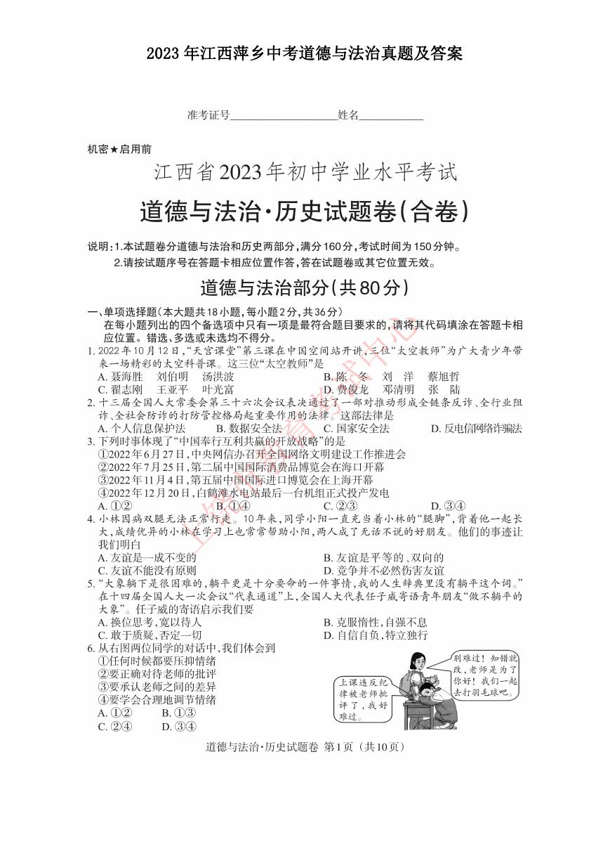 2023年江西萍乡中考道德与法治真题及答案.doc
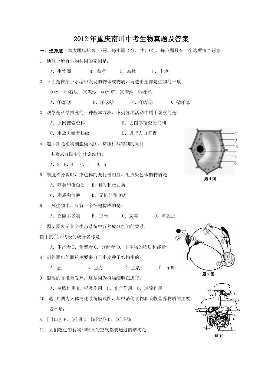 2012年重庆南川中考生物真题及答案.doc
 2013年江西师范大学地理学综合及文艺理论基础考研真题.doc
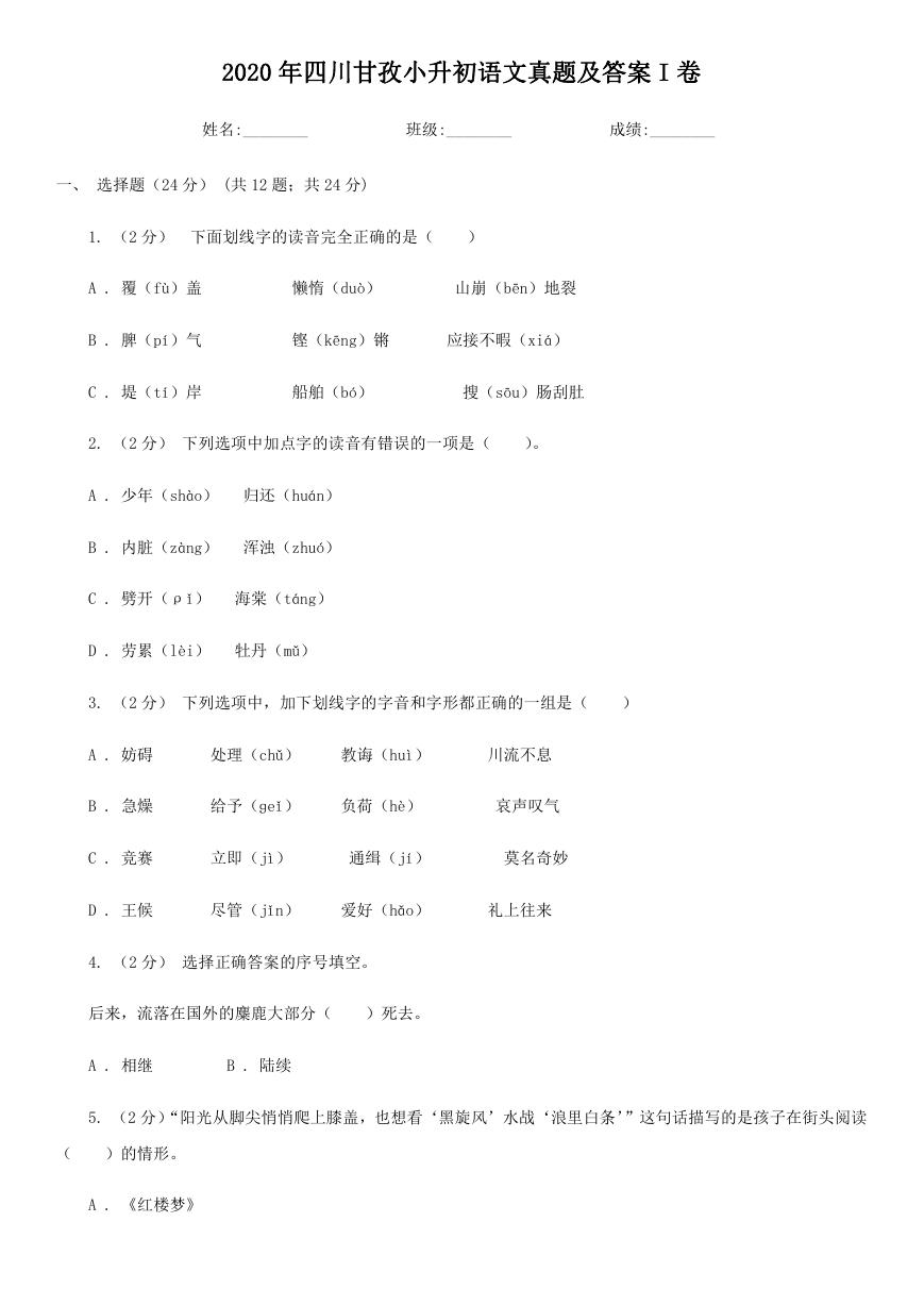 2020年四川甘孜小升初语文真题及答案I卷.doc
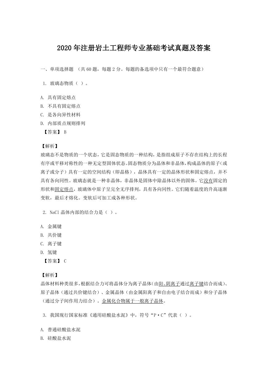 2020年注册岩土工程师专业基础考试真题及答案.doc
 2023-2024学年福建省厦门市九年级上学期数学月考试题及答案.doc
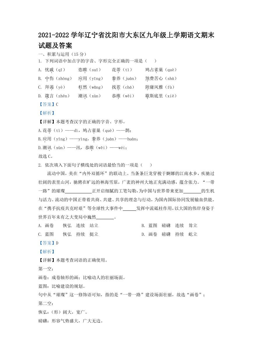 2021-2022学年辽宁省沈阳市大东区九年级上学期语文期末试题及答案.doc
 2022-2023学年北京东城区初三第一学期物理期末试卷及答案.doc
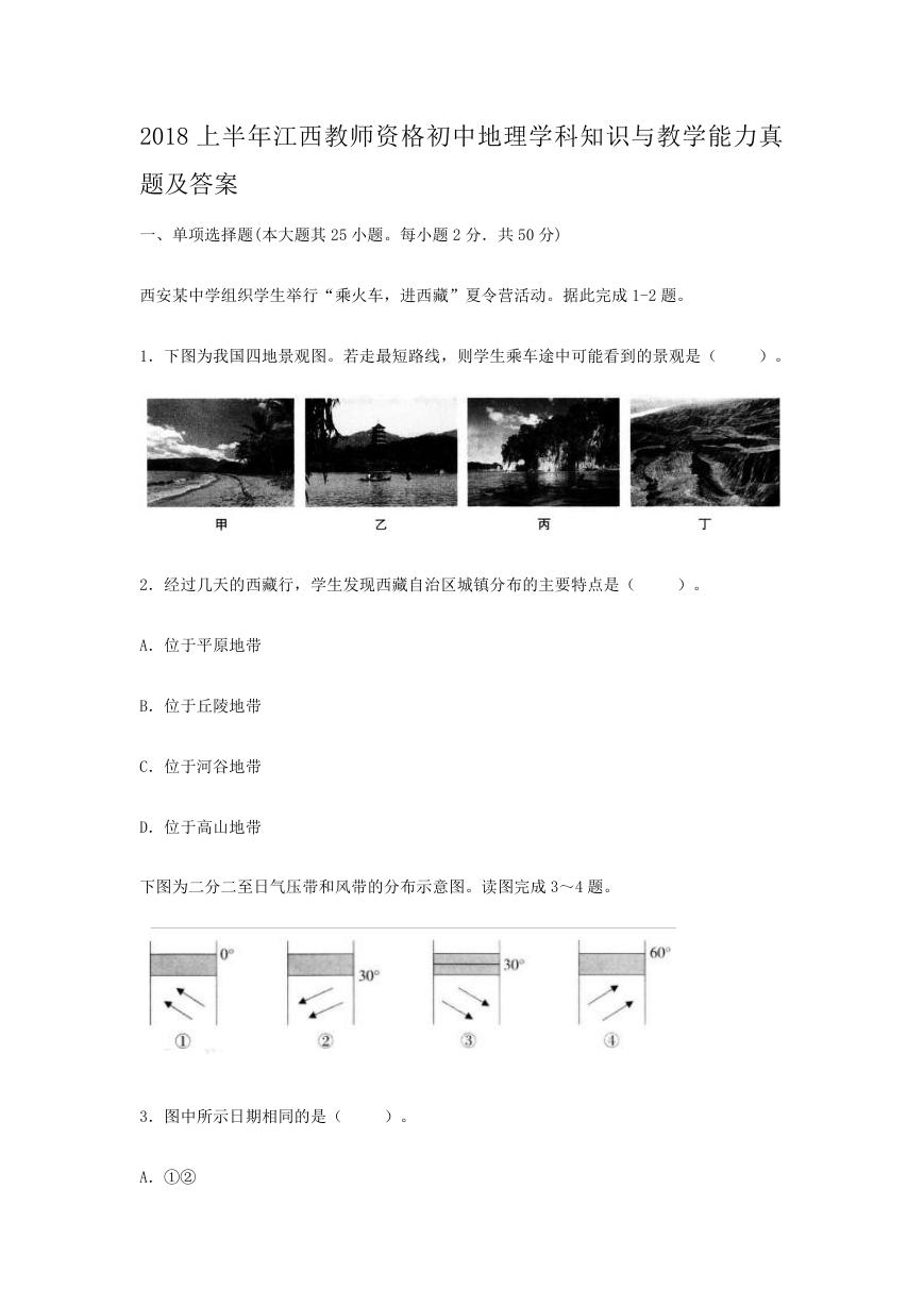 2018上半年江西教师资格初中地理学科知识与教学能力真题及答案.doc
 2012年河北国家公务员申论考试真题及答案-省级.doc
 2020-2021学年江苏省扬州市江都区邵樊片九年级上学期数学第一次质量检测试题及答案.doc
 2022下半年黑龙江教师资格证中学综合素质真题及答案.doc