�
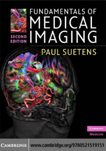
Fundamentals of Medical Imaging
Second Edition
�
Fundamentalsof
Medical Imaging
Second Edition
Paul Suetens
Katholieke Universiteit Leuven
�
CAMBRIDGE UNIVERSITY PRESS
Cambridge, New York, Melbourne, Madrid, Cape Town, Singapore,
São Paulo, Delhi, Dubai, Tokyo
Cambridge University Press
The Edinburgh Building, Cambridge CB2 8RU, UK
Published in the United States of America by Cambridge University Press, New York
www.cambridge.org
Information on this title: www.cambridge.org/9780521519151
© First edition © Cambridge University Press 2002
Second edition © P. Suetens 2009
This publication is in copyright. Subject to statutory exception and to the
provision of relevant collective licensing agreements, no reproduction of any part
may take place without the written permission of Cambridge University Press.
First published in print format
2009
ISBN-13 978-0-511-59640-7
eBook (NetLibrary)
ISBN-13 978-0-521-51915-1
Hardback
Cambridge University Press has no responsibility for the persistence or accuracy
of urls for external or third-party internet websites referred to in this publication,
and does not guarantee that any content on such websites is, or will remain,
accurate or appropriate.
Every effort has been made in preparing this publication to provide accurate and
up-to-date information which is in accord with accepted standards and practice at
the time of publication. Although case histories are drawn from actual cases, every
effort has been made to disguise the identities of the individuals involved.
Nevertheless, the authors, editors and publishers can make no
warranties that the information contained herein is totally free from error, not least
because clinical standards are constantly changing through research and
regulation. The authors, editors and publishers therefore disclaim all liability for
direct or consequential damages resulting from the use of material contained in
this publication. Readers are strongly advised to pay careful attention to
information provided by the manufacturer of any drugs or equipment that they
plan to use.
�
Contents
page vii
Preface
Acknowledgments
ix
1 Introduction to digital image
1
processing
Digital images
Image quality
Basic image operations
1
2
4
16
14
14
2 Radiography
Introduction
X-rays
14
Interaction with matter
X-ray detectors
17
Dual-energy imaging
Image quality
Equipment
Clinical use
Biologic effects and safety
Future expectations
32
24
25
23
21
27
59
3 X-ray computed tomography
34
35
33
Introduction
X-ray detectors in CT
Imaging
Cardiac CT
46
Dual-energy CT
Image quality
Equipment
Clinical use
Biologic effects and safety
Future expectations
63
53
58
49
48
4 Magnetic resonance imaging
64
Introduction
Physics of the transmitted signal
Interaction with tissue
68
Signal detection and detector
Imaging
72
71
33
64
64
90
Image quality
Equipment
Clinical use
Biologic effects and safety
104
Future expectations
95
98
102
5 Nuclear medicine imaging
105
Introduction
Radionuclides
Interaction of γ-photons and particles
105
105
with matter
108
116
108
Data acquisition
Imaging
111
Image quality
Equipment
Clinical use
Biologic effects and safety
Future expectations
125
117
122
6 Ultrasound imaging
128
Introduction
Physics of acoustic waves
Generation and detection of
128
ultrasound
137
145
141
138
Gray scale imaging
Doppler imaging
Image quality
Equipment
Clinical use
Biologic effects and safety
Future expectations
156
149
152
124
128
155
7 Medical image analysis
159
159
Introduction
Manual analysis
Automated analysis
Computational strategies for automated
160
160
medical image analysis
166
Pixel classification
163
�
Contents
Geometric model matching using a
transformation matrix
170
Flexible geometric model matching
Validation
Future expectations
189
186
8 Visualization for diagnosis
and therapy
Introduction
2D visualization
3D rendering
190
190
192
192
175
205
Virtual reality
User interaction
Intraoperative navigation
214
Augmented reality
Future expectations
218
207
208
Appendix A: Linear system theory
Appendix B: Exercises
Bibliography
248
Index
232
246
219
vi
�
Preface
This book explains the applied mathematical and
physical principles of medical imaging and image pro-
cessing. It gives a complete survey, accompanied by
more than 300 illustrations in color, of how medical
images are obtained and how they can be used for
diagnosis, therapy, and surgery.
It has been written principally as a course text on
medical imaging intended for graduate and final-year
undergraduate students with a background in physics,
mathematics, or engineering. However, I have made
an effort to make the textbook readable for biomedical
scientists and medical practitioners as well by delet-
ing unnecessary mathematical details, without giving
up the depth needed for physicists and engineers.
Mathematical proofs are highlighted in separate para-
graphs and can be skipped without hampering a fluent
reading of the text.
Although a large proportion of the book covers
the physical principles of imaging modalities,
the
emphasis is always on how the image is computed.
Equipment design, clinical considerations, and diag-
nosis are treated in less detail. Premature techniques
or topics under investigation have been omitted.
Presently, books on medical imaging fall into two
groups, neither of which is suitable for this read-
ership. The first group is the larger and comprises
books directed primarily at the less numerate pro-
fessions such as physicians, surgeons, and radiologic
technicians. These books cover the physics and mathe-
matics of all the major medical imaging modalities, but
mostly in a superficial way. They do not allow any real
understanding of these imaging modalities. The sec-
ond group comprises books suitable for professional
medical physicists or researchers with expertise in the
field. Although these books have a numerate approach,
they tend to cover the topics too deeply for the
beginner and to have a narrower scope than this book.
The text reflects what I teach in class, but there is
somewhat more material than I can cover in a module
of 30 contact hours. This means that there is scope for
the stronger student to read around the subject and
also makes the book a useful purchase for those going
on to do research.
In Chapter 1, an introduction to digital image pro-
cessing is given. It summarizes the jargon used by
the digital image community, the components defin-
ing image quality, and basic image operations used
to process digital images. The theory of linear sys-
tems, described in Chapter 2 of the first edition, has
been moved to an appendix. It is too high-level for
the medical reader and a significant part of the engi-
neering readers of the previous edition considered it
as redundant. However, many students in physics or
engineering are not familiar with linear system theory
and will welcome this appendix.
Chapters 2–6 explain how medical images are
obtained. The most important imaging modalities
today are discussed: radiography, computed tomogra-
phy, magnetic resonance imaging, nuclear medicine
imaging,
and ultrasonic imaging. Each chapter
includes (1) a short history of the imaging modality,
(2) the theory of the physics of the signal and its inter-
action with tissue, (3) the image formation or recon-
struction process, (4) a discussion of the image quality,
(5) the different types of equipment in use today, (6)
examples of the clinical use of the modality, (7) a brief
description of the biologic effects and safety issues, and
(8) some future expectations. The imaging modalities
have made an impressive evolution in a short time with
respect to quality, size and applicability. This part of
the book provides up-to-date information about these
systems.
Chapters 7 and 8 deal with image analysis and
visualization for diagnosis, therapy and surgery once
images are available. Medical images can, for example,
be analyzed to obtain quantitative data, or they can
be displayed in three dimensions and actively used
to guide a surgical intervention. Most courses sepa-
rate the imaging theory from the postprocessing, but
I strongly believe that they should be taken together
�




 2023年江西萍乡中考道德与法治真题及答案.doc
 2012年重庆南川中考生物真题及答案.doc
 2013年江西师范大学地理学综合及文艺理论基础考研真题.doc
 2020年四川甘孜小升初语文真题及答案I卷.doc
 2020年注册岩土工程师专业基础考试真题及答案.doc
 2023-2024学年福建省厦门市九年级上学期数学月考试题及答案.doc
 2021-2022学年辽宁省沈阳市大东区九年级上学期语文期末试题及答案.doc
 2022-2023学年北京东城区初三第一学期物理期末试卷及答案.doc
 2018上半年江西教师资格初中地理学科知识与教学能力真题及答案.doc
 2012年河北国家公务员申论考试真题及答案-省级.doc
 2020-2021学年江苏省扬州市江都区邵樊片九年级上学期数学第一次质量检测试题及答案.doc
 2022下半年黑龙江教师资格证中学综合素质真题及答案.doc