DRAFT INTERNATIONAL STANDARD
ISO/TC 229
Voting begins on:
Secretariat: BSI
Voting terminates on:
ISO/DIS 19749
2018-08-30
2018-11-22
Nanotechnologies — Measurements of particle size and
shape distributions by scanning electron microscopy
Nanotechnologies — Détermination de la taille et de la distribution en taille des nano-objets par
microscopie électronique à balayage
ICS: 07.120
THIS DOCUMENT IS A DRAFT CIRCULATED
FOR COMMENT AND APPROVAL.
IT
IS
THEREFORE SUBJECT TO CHANGE AND MAY
NOT BE REFERRED TO AS AN INTERNATIONAL
STANDARD UNTIL PUBLISHED AS SUCH.
IN ADDITION TO THEIR EVALUATION AS
BEING ACCEPTABLE
INDUSTRIAL,
FOR
TECHNOLOGICAL,
COMMERCIAL
AND
USER PURPOSES, DRAFT
INTERNATIONAL
STANDARDS MAY ON OCCASION HAVE TO
BE CONSIDERED IN THE LIGHT OF THEIR
POTENTIAL TO BECOME STANDARDS TO
WHICH REFERENCE MAY BE MADE
IN
NATIONAL REGULATIONS.
RECIPIENTS OF THIS DRAFT ARE INVITED
TO SUBMIT, WITH THEIR COMMENTS,
NOTIFICATION OF ANY RELEVANT PATENT
RIGHTS OF WHICH THEY ARE AWARE AND TO
PROVIDE SUPPORTING DOCUMENTATION.
This document is circulated as received from the committee secretariat.
Reference number
ISO/DIS 19749:2018(E)
© ISO 2018
�
ISO/DIS 19749:2018(E)
COPYRIGHT PROTECTED DOCUMENT
© ISO 2018
All rights reserved. Unless otherwise specified, or required in the context of its implementation, no part of this publication may
be reproduced or utilized otherwise in any form or by any means, electronic or mechanical, including photocopying, or posting
on the internet or an intranet, without prior written permission. Permission can be requested from either ISO at the address
below or ISO’s member body in the country of the requester.
ISO copyright office
CP 401 • Ch. de Blandonnet 8
CH-1214 Vernier, Geneva
Phone: +41 22 749 01 11
Fax: +41 22 749 09 47
Email: copyright@iso.org
Website: www.iso.org
Published in Switzerland
ii
© ISO 2018 – All rights reserved
�
ISO/DIS 19749:2018(E)
1
2
3
Contents
Page
Foreword ..........................................................................................................................................................................................................................................v
Introduction ................................................................................................................................................................................................................................vi
Scope .................................................................................................................................................................................................................................1
Normative references ......................................................................................................................................................................................1
Terms and definitions .....................................................................................................................................................................................2
General ...........................................................................................................................................................................................................2
3.1
Core terms: image analysis ...........................................................................................................................................................4
3.2
3.3
Core terms: statistical symbols and definitions .........................................................................................................5
3.4
Core terms: measurands and descriptors ........................................................................................................................8
Symbols ....................................................................................................................................................................................8
3.4.1
3.4.2
Terms .........................................................................................................................................................................................9
3.5
Core terms: metrology ...................................................................................................................................................................12
3.6
Core terms: scanning electron microscopy .................................................................................................................14
General principles ............................................................................................................................................................................................16
SEM imaging ...........................................................................................................................................................................................16
4.1
4.2
SEM image-based particle size measurements ........................................................................................................16
SEM image-based particle shape measurements ...................................................................................................18
4.3
Sample preparation ........................................................................................................................................................................................19
General recommendations.........................................................................................................................................................19
5.1
5.2
Ensuring good sampling of powder or dispersion-in-liquid raw materials ....................................19
5.3
Ensuring representative dispersion ..................................................................................................................................20
5.4
Nanoparticle deposition on a substrate .........................................................................................................................20
5.4.1 Nanoparticle deposition on silicon or other wafers and chips .............................................21
5.4.2 Nanoparticle deposition on TEM grids ......................................................................................................22
5.5
The number of samples to be prepared .........................................................................................................................23
5.6
The number of particles to be measured for particle size determination ........................................23
5.7
The number of particles to be measured for particle shape determination ...................................23
Qualification of the SEM .............................................................................................................................................................................24
6.1
General ........................................................................................................................................................................................................24
6.2 Measurement of spatial resolution .....................................................................................................................................24
6.3 Measurement of drifts ...................................................................................................................................................................24
6.4 Measurement of electron beam-induced contamination ................................................................................25
6.5 Measurement of scale and linearity ...................................................................................................................................26
6.6 Measurement of noise ...................................................................................................................................................................26
6.7 Measurements of primary electron beam current ................................................................................................27
Image acquisition .............................................................................................................................................................................................27
7.1
Setting suitable image magnification and pixel resolution ............................................................................32
Particle analysis .................................................................................................................................................................................................33
8.1
Individual particle analysis .......................................................................................................................................................33
8.2
Automated particle analysis .....................................................................................................................................................33
8.3
Automated particle analysis procedure example ...................................................................................................34
Data analysis ..........................................................................................................................................................................................................34
9.1
Raw data screening: detecting touching particles, artifacts and contaminants ..........................35
9.2
Fitting models to data ....................................................................................................................................................................35
Assessment of measurement uncertainty ....................................................................................................................35
9.3
9.3.1
Example: measurement uncertainty for particle size measurements ............................36
9.3.2
Bivariate analysis ..........................................................................................................................................................36
Reporting the results ....................................................................................................................................................................................37
Annex A (normative) Uncertainty measurement in relation to spatial resolution of the SEM .............38
iii
© ISO 2018 – All rights reserved
7
8
10
4
5
6
9
�
ISO/DIS 19749:2018(E)
Annex B (informative) Cross-sectional titanium dioxide samples preparation ...................................................39
Annex C (informative) Case study on well dispersed 60 nm size silicon dioxide nanoparticles .........41
Annex D (informative) Case study on 40 nm size titanium dioxide nanoparticles ...........................................48
Annex E (informative) Example for extracting particle size results of SEM-based
nanoparticle measurements using Image J ...........................................................................................................................56
Annex F (informative) The effects of some image acquisition parameters and thresholding
methods on SEM particle size measurements ....................................................................................................................59
Annex G (informative) Example for reporting results of SEM-based nanoparticle measurements 62
Bibliography .............................................................................................................................................................................................................................71
iv
© ISO 2018 – All rights reserved
�
ISO/DIS 19749:2018(E)
Foreword
ISO (the International Organization for Standardization) is a worldwide federation of national standards
bodies (ISO member bodies). The work of preparing International Standards is normally carried out
through ISO technical committees. Each member body interested in a subject for which a technical
committee has been established has the right to be represented on that committee. International
organizations, governmental and non-governmental, in liaison with ISO, also take part in the work.
ISO collaborates closely with the International Electrotechnical Commission (IEC) on all matters of
electrotechnical standardization.
The procedures used to develop this document and those intended for its further maintenance are
described in the ISO/IEC Directives, Part 1. In particular the different approval criteria needed for the
different types of ISO documents should be noted. This document was drafted in accordance with the
editorial rules of the ISO/IEC Directives, Part 2 (see www .iso .org/directives).
Attention is drawn to the possibility that some of the elements of this document may be the subject of
patent rights. ISO shall not be held responsible for identifying any or all such patent rights. Details of
any patent rights identified during the development of the document will be in the Introduction and/or
on the ISO list of patent declarations received (see www .iso .org/patents).
Any trade name used in this document is information given for the convenience of users and does not
constitute an endorsement.
For an explanation on the meaning of ISO specific terms and expressions related to conformity assessment,
as well as information about ISO's adherence to the World Trade Organization (WTO) principles in the
Technical Barriers to Trade (TBT) see the following URL: www .iso .org/iso/foreword .html.
The committee responsible for this document is ISO/TC 229 Nanotechnologies.
© ISO 2018 – All rights reserved
v
�
ISO/DIS 19749:2018(E)
Introduction
This document provides guidance for measuring and reporting the size and shape distributions of
nanometer-scale particles using images acquired by the scanning electron microscope (SEM). This
document applies to the SEM-based measurement of larger particles also. Nanoparticles are three-
dimensional (3D) objects, but the SEM image is only a two-dimensional (2D) representation of the 3D
shape from a certain viewing angle. The SEM image carries valuable information about the size and
shape of particles. While the SEM image does contain a certain amount of 3D information, for sake of
simplicity, this document does not deal with reconstructing 3D information. Rigorous three-dimensional
characterization of nanoparticles would include size, shape, surface structure (e.g. texture), surface
and internal material composition, and their locations in the investigated 3D volume. This document
deals with two attributes of morphology, size and shape, for discrete and aggregated nano-objects
(materials with at least one dimension in the nanometer-scale, i.e., within 1 nm to 100 nm). Suitable
sample preparation is essential to obtaining high-quality electron microscope images and preferred
techniques often vary with the sample material. It is equally important to make sure that the SEM itself
is suitable to carry out the measurements with the required uncertainty. Typical guidance suggests that
a large number, several hundreds or thousands of particles need to be measured for statistically sound
size and shape distribution results. The actual number of nano-objects needed to be measured depends
on the sample, the required uncertainty and on the performance of the SEM. Statistical evaluation of
the data and the evaluation of uncertainty of the measurands are included as part of the measurement
and reporting procedures.
This document contains measurement procedures, particle and data analysis and reporting sections.
In Annexes, there are specific examples for measurements and guidance for the qualification of the
SEM for reliable quantitative measurements. Automation of the image acquisition and data analysis can
reduce cost and improve the quality of the results. Measurements of samples of discrete nanoparticles
are generally easier to carry out with automated image acquisition and particle analysis systems.
Measurements of complex discrete nanoparticles, and aggregates or agglomerates of nanoparticles
may require operator-assisted image acquisition and analysis.
vi
© ISO 2018 – All rights reserved
�
DRAFT INTERNATIONAL STANDARD
ISO/DIS 19749:2018(E)
Nanotechnologies — Measurements of particle size and
shape distributions by scanning electron microscopy
1 Scope
2 Normative references
This International Standard specifies methods of determining nanoparticle size and shape distributions
by acquiring and evaluating scanning electron microscope images and by obtaining and reporting
accurate results.
NOTE 1
This International Standard applies to particles with a lower size limit that depends on the
required uncertainty and on the suitable performance of the SEM, which must be proven first -according to the
requirements described in this document.
This International Standard applies also to SEM-based size and shape measurements of larger than
NOTE 2
nanoscale particles.
The following referenced documents are indispensable for the application of this document. For dated
references, only the edition cited applies. For undated references, the latest editions of the referenced
documents (including any amendments) apply.
ISO 13322-1:2014, Particle size analysis — Image analysis methods — Part 1: Static image analysis
methods [1]
ISO 5725-1:1994, Accuracy (trueness and precision) of measurement methods and results — Part 1: General
principles and definitions [2]
ISO 9276-1:1998, Representation of results of particle size analysis — Part 1: Graphical representation [3]
ISO 9276-2:2014, Representation of results of particle size analysis — Part 2: Calculation of average
particle sizes/diameters and moments from particle size distributions [4]
ISO 9276-3:2008, Representation of results of particle size analysis — Part 3: Adjustment of an experimental
curve to a reference model [5]
ISO 9276-5:2005, Representation of results of particle size analysis — Part 5: Methods of calculation
relating to particle size analyses using logarithmic normal probability distribution [6]
ISO 9276-6:2008, Representation of results of particle size analysis — Part 6: Descriptive and quantitative
representation of particle shape and morphology [7]. This document provides a detailed list of other
shape parameters for size, macroshape descriptors, geometrical descriptors, proportion descriptors,
and mesoshape descriptors.
ISO 26824:2013, Particle characterization of particulate systems — Vocabulary [8]
ISO 29301:2017, Microbeam analysis — Analytical electron microscopy — Methods for calibrating image
magnification by using reference materials with periodic structures [9]
ISO/TS 80004-1:2015, Nanotechnologies — Vocabulary — Part 1: Core terms [10]
ISO/TS 80004-2:2015, Nanotechnologies — Vocabulary — Part 2: Nano-objects [11]
ISO/TS 80004-3:2010, Nanotechnologies — Vocabulary — Part 3: Carbon nano-objects [12]
ISO/TS 80004-4:2011, Nanotechnologies — Vocabulary — Part 4: Nanostructured materials [13]
© ISO 2018 – All rights reserved
1
�
ISO/DIS 19749:2018(E)
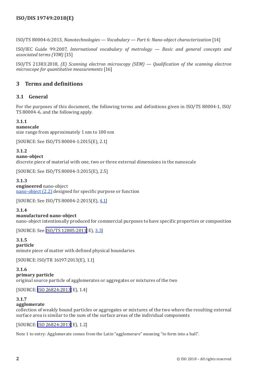
3.1 General
3.1.1
nanoscale
3.1.2
nano-object
3.1.3
3.1.5
particle
3 Terms and definitions
ISO/TS 80004-6:2013, Nanotechnologies — Vocabulary — Part 6: Nano-object characterization [14]
ISO/IEC Guide 99:2007, International vocabulary of metrology — Basic and general concepts and
associated terms (VIM) [15]
ISO/TS 21383:2018, (E) Scanning electron microscopy (SEM) — Qualification of the scanning electron
microscope for quantitative measurements [16]
For the purposes of this document, the following terms and definitions given in ISO/TS 80004-1, ISO/
TS 80004-6, and the following apply.
size range from approximately 1 nm to 100 nm
[SOURCE: See ISO/TS 80004-1:2015(E), 2.1]
discrete piece of material with one, two or three external dimensions in the nanoscale
[SOURCE: See ISO/TS 80004-3:2015(E), 2.5]
engineered nano-object
nano-object (2.2) designed for specific purpose or function
[SOURCE: See ISO/TS 80004-2:2015(E), 4.1]
nano-object intentionally produced for commercial purposes to have specific properties or composition
[SOURCE: See ISO/TS 12805:2011(E), 3.3]
minute piece of matter with defined physical boundaries
[SOURCE: ISO/TR 16197:2013(E), 1.1]
original source particle of agglomerates or aggregates or mixtures of the two
[SOURCE: ISO 26824:2013(E), 1.4]
collection of weakly bound particles or aggregates or mixtures of the two where the resulting external
surface area is similar to the sum of the surface areas of the individual components
[SOURCE: ISO 26824:2013(E), 1.2]
Note 1 to entry: Agglomerate comes from the Latin “agglomerare” meaning “to form into a ball”.
3.1.4
manufactured nano-object
3.1.6
primary particle
3.1.7
agglomerate
2
© ISO 2018 – All rights reserved
�





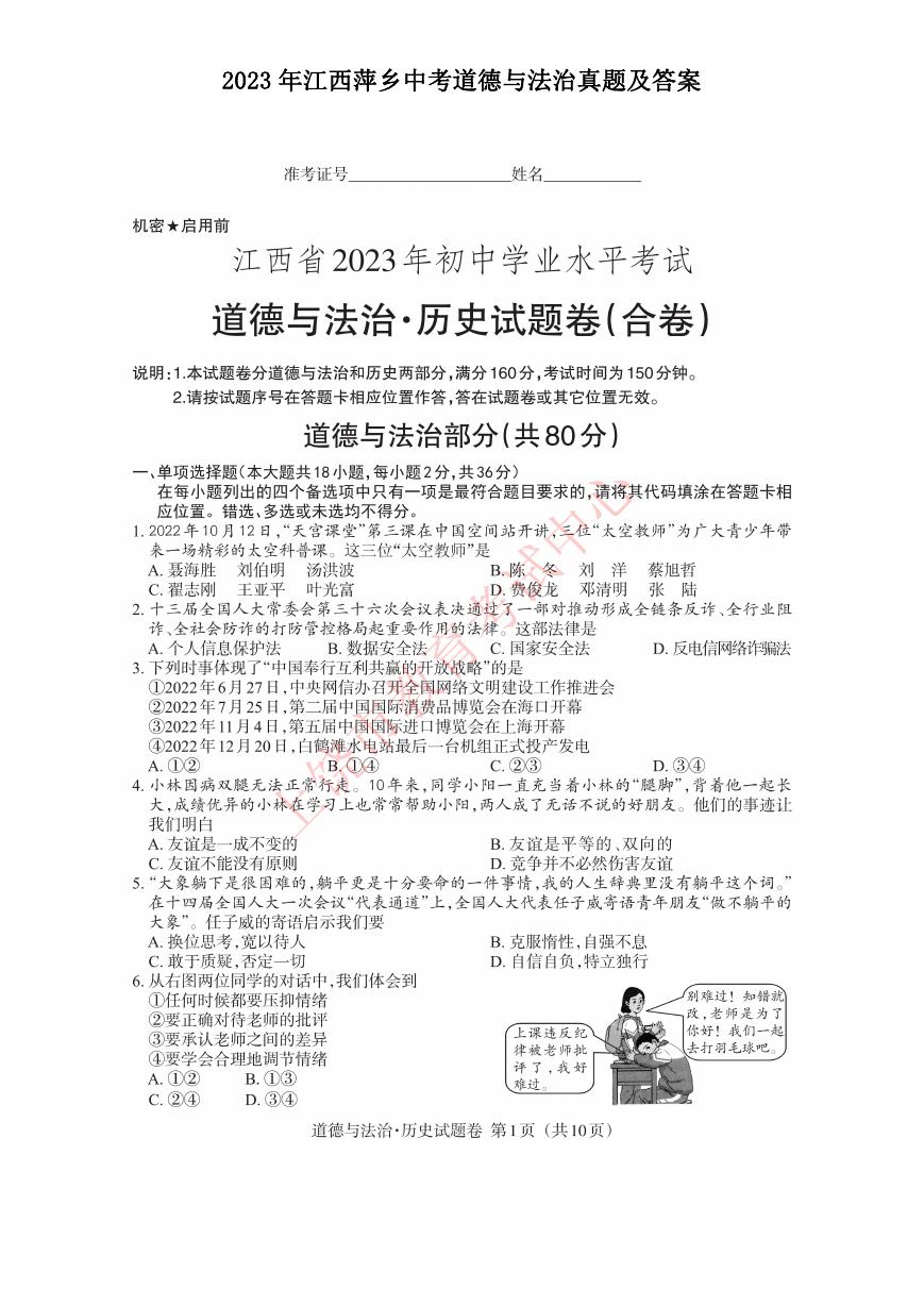 2023年江西萍乡中考道德与法治真题及答案.doc
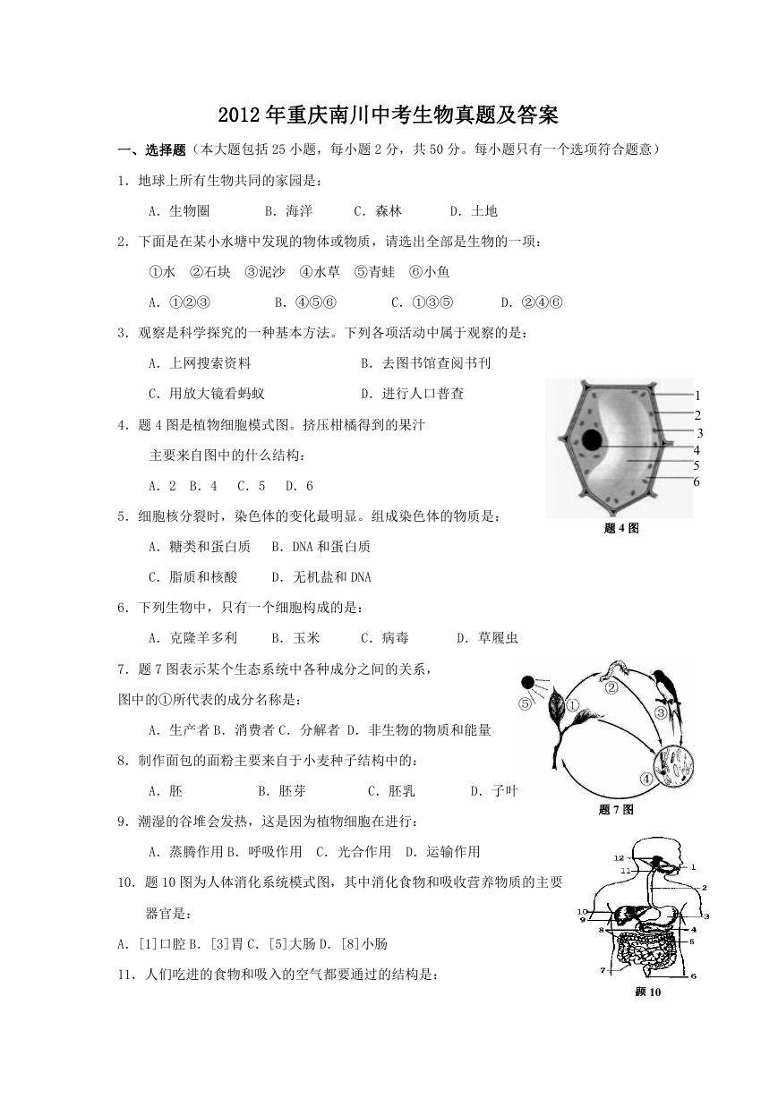 2012年重庆南川中考生物真题及答案.doc
 2013年江西师范大学地理学综合及文艺理论基础考研真题.doc
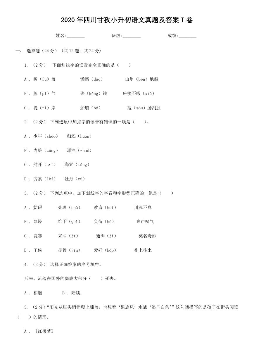 2020年四川甘孜小升初语文真题及答案I卷.doc
 2020年注册岩土工程师专业基础考试真题及答案.doc
 2023-2024学年福建省厦门市九年级上学期数学月考试题及答案.doc
 2021-2022学年辽宁省沈阳市大东区九年级上学期语文期末试题及答案.doc
 2022-2023学年北京东城区初三第一学期物理期末试卷及答案.doc
 2018上半年江西教师资格初中地理学科知识与教学能力真题及答案.doc
 2012年河北国家公务员申论考试真题及答案-省级.doc
 2020-2021学年江苏省扬州市江都区邵樊片九年级上学期数学第一次质量检测试题及答案.doc
 2022下半年黑龙江教师资格证中学综合素质真题及答案.doc