Journal of Modern Physics, 2018, 9, 2274-2285
http://www.scirp.org/journal/jmp
ISSN Online: 2153-120X
ISSN Print: 2153-1196
Evaluation of Deformable Image Registration
and Dose Accumulation Using Histogram
Matching Algorithm between kVCT and MVCT
with Helical Tomotherapy
Masahide Saito*, Yuki Shibata, Naoki Sano, Kengo Kuriyama, Takafumi Komiyama,
Kan Marino, Shinichi Aoki, Hiroshi Onishi
Department of Radiology, University of Yamanashi, Yamanashi, Japan
How to cite this paper: Saito, M., Shibata,
Y., Sano, N., Kuriyama, K., Komiyama, T.,
Marino, K., Aoki, S. and Onishi, H. (2018)
Evaluation of Deformable Image Registra-
tion and Dose Accumulation Using Histo-
gram Matching Algorithm between kVCT
and MVCT with Helical Tomotherapy.
Journal of Modern Physics, 9, 2274-2285.
https://doi.org/10.4236/jmp.2018.913143
Received: October 8, 2018
Accepted: October 30, 2018
Published: November 2, 2018
Copyright © 2018 by authors and
Scientific Research Publishing Inc.
This work is licensed under the Creative
Commons Attribution International
License (CC BY 4.0).
http://creativecommons.org/licenses/by/4.0/
Open Access
Abstract
Purpose: To evaluate the accuracy of deformable image registration (DIR)
between the planning kVCT (pCT) and the daily MVCT combined with the
histogram matching (HM) algorithm, and evaluate the deformable dose ac-
cumulation using a suggested method for adaptive radiotherapy with Helical
Tomotharapy (HT). Methods: For five prostate cancer patients (76 Gy/38 Fr)
treated with HT in our institution, seven MVCT series (a total of 35 series)
acquired weekly were investigated. First, to minimize the effect of different
HU values between pCT and MVCT, this image-processing method adjusts
HU values between pCT and MVCT images by using image cumulative his-
tograms of HU values, generating an HM-MVCT. Then, the DIR of the pCT
to the HM-MVCT was performed, generating a deformed pCT. Finally, de-
formable dose accumulation was performed toward the pCT image. Results:
The accuracy of DIR was significantly improved by using the HM algorithm,
compared with non-HM method for several structures (p < 0.05). The mean
dice similarity coefficient of the non-HM method was 0.75 ± 0.05, 0.83 ± 0.06,
and 0.90 ± 0.04 for the CTV, rectum, and bladder, respectively, while that of
the HM method was 0.81 ± 0.06, 0.81 ± 0.04, and 0.92 ± 0.06, respectively.
For the deformable dose accumulation, some difference was observed be-
tween the two methods, particularly for the small calculated regions, such as
rectum V60 and V70. Conclusion: Adapting the HM method can improve
the accuracy of DIR. Furthermore, dose calculation using the deformed pCT
using HM methods can be an effective tool for adaptive radiotherapy.
Keywords
Radiotherapy, Tomotherapy, MVCT, Histogram-Matching, Deformable Image
DOI: 10.4236/jmp.2018.913143 Nov. 2, 2018
2274
Journal of Modern Physics
�
M. Saito et al.
Registration
1. Introduction
Intensity modulated radiotherapy (IMRT) using helical tomotherapy (HT) (Ac-
curay, Sunnyvale, CA) reduces the radiation dose to organs at risks (OAR) while
maintaining conformity of the tumor [1]. Meanwhile, image-guided radiothera-
py using HT uses mega-voltage computed tomography (MVCT) images [2],
which have been used routinely for patient setup in HT treatments. Under-
standing daily anatomical changes and calculating the radiation dose are imper-
ative. Therefore, some studies on adaptive radiotherapy using the daily MVCT
and deformable image registration (DIR) have been published [3] [4] [5] [6].
Recently, Branchini et al. suggested a method of planning CT (pCT) for daily
MVCT deformation and evaluated its accuracy [7]. They suggested that the me-
thod might be sufficiently fast and reliable for daily delivered dose evaluations in
clinical strategies for adaptive radiotherapy using HT. However, the use of the
daily MVCT is generally limited due to significant photoelectric interaction
components and the presence of noise resulting from low detector quantum effi-
ciency of megavoltage x-rays. The poor image quality of MVCT might decrease
the accuracy of DIR and dose calculation. Although several authors introduced
denoising methods [3], further evaluation for more accurate ART methods using
MVCT is needed.
Some authors have investigated the improvement of image quality using
Hounsfield unit (HU) modification methods, such as histogram matching (HM)
method [8] [9] [10]. Although previous studies investigated kV-CBCT, no re-
ports on improving the quality of MVCT images have been published. Further-
more, the deformable dose accumulation using this method was not investigated
sufficiently. Therefore, the purpose of this study was to evaluate the accuracy of
DIR between the pCT and the daily MVCT combined with the HM algorithm.
In addition, we evaluated the deformable dose accumulation using a suggested
method for adaptive radiotherapy with HT.
2. Materials and Methods
2.1. Planning kVCT and MVCT Images
For five prostate cancer patients (76 Gy/38 Fr) treated with HT in our institu-
tion, seven MVCT series (a total of 35 series) acquired weekly were investigated.
These images were used for routine patient setup and acquired with the pitch of
4 mm and reconstruction thickness of 2 mm. For the pCT, the settings for image
acquisition were 120 kV, 250 mA, 0.50 s, and a 2.0 mm slice thickness.
For the pCT image and each MVCT image, anatomical contours of the CTV
(prostate + seminal vesicle), rectum, and bladder were described by an experi-
mental oncologist and medical physicist. However, the scan range of each MVCT
2275
Journal of Modern Physics
DOI: 10.4236/jmp.2018.913143
�
M. Saito et al.
was insufficient to describe the whole anatomical volume of the rectum and
bladder in some patients.
2.2. DIR Using Histogram Matching Algorithm
In this study, the scan range of the daily MVCT was smaller than the pCT as
mentioned previously. To perform DIR between pCT and MVCT normally, we
generated a merged MVCT, as shown in Figure 1. First, the rigid image registra-
tion was performed between pCT and each MVCT image. Then, the image re-
gion outside the MVCT was replaced with pCT, resulting in the merged MVCT.
The algorithm between the pCT and the fractional MVCT was applied to im-
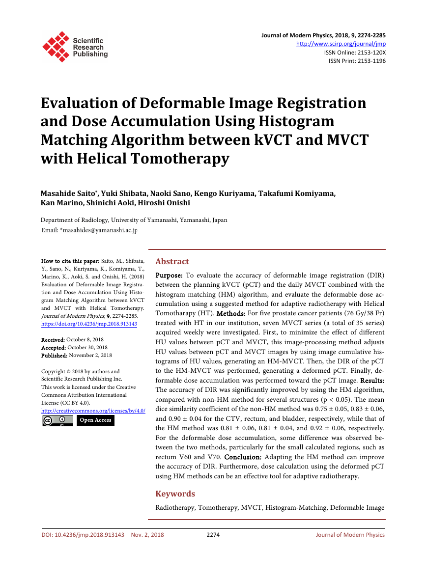
prove the accuracy of pCT to MVCT deformation. Figure 2(a) shows the pro-
cedure of the HM method. To minimize the effect of different HU values be-
tween pCT and MVCT, this image-processing method adjusts HU values be-
tween pCT and MVCT images by using image cumulative histograms of HU
values, generating an HM-MVCT. The HM method was performed by a script
generated using MATLAB 2016a (Mathworks Inc., Natick, MA).
Then, the DIR of the pCT to the HM-MVCT was performed, generating a de-
formed pCT, as shown as Figure 2(b). In this study, this method was also ap-
plied to the original MVCT and the result was compared with that of the HM
method. All DIRs were performed using the MIM Maestro ver.6.6.8 (MIM Soft-
ware, Inc., Cleveland, OH) with intensity-based free form deforamtion algo-
rithm.
2.3. Dose Accumulation
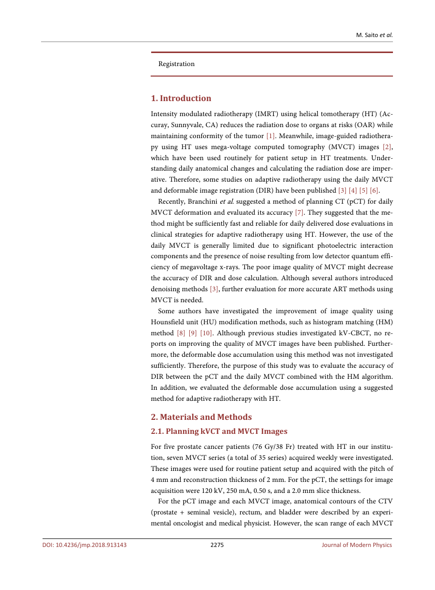
Figure 3 demonstrates the workflow of the deformable dose accumulation in
Figure 1. Concept of a merged MVCT. First, the rigid image registration was performed
between the pCT and each fractional MVCT image. Then, the image region outside the
MVCT was replaced with that of the pCT.
2276
Journal of Modern Physics
DOI: 10.4236/jmp.2018.913143
�
M. Saito et al.
Figure 2. The procedure of deformable image registration combined with the HM me-
thod. First, to minimize the effect of different HU values between pCT and MVCT, this
image-processing method adjusts HU values between pCT and MVCT images by using
image cumulative histograms of HU values, generating an HM-MVCT (a). Then, the DIR
of the pCT to the HM-MVCT was performed, generating a deformed pCT (b).
Figure 3. The workflow of the deformable dose accumulation in this study. First, each
fractional deformed pCT was generated by using HM-MVCT (or non HM-MVCT).
Then, the dose of the weekly deformed pCT was calculated. Finally, the deformable dose
accumulation was performed toward the pCT image.
2277
Journal of Modern Physics
DOI: 10.4236/jmp.2018.913143
�
M. Saito et al.
this study. First, each fractional deformed pCT was generated by using
HM-MVCT (or non HM-MVCT). Then, the dose of the weekly deformed pCT
was calculated as following condition. The target prescribed dose that was calcu-
lated by using each weekly MVCT image was scaled to 10.86 Gy, which was di-
vided by the total dose (76 Gy) by 7 weeks. Finally, the deformable dose accu-
mulation was performed toward the pCT image using MIM software. The dose
calculation of the reference pCT image was performed using Tomotherapy plan-
ning station ver. 5.1.0.2, and each fractional dose of the deformed pCT was cal-
culated via Tomotherapy DQA station ver. 5.1.0.4 with the fine condition of
dose grid size.
2.4. Evaluation and Analysis
To evaluate the accuracy of the DIR, dice similarity coefficients (DSC) between
deformed contours described in deformed pCT and manual contours described
in original MVCT were calculated for CTV, rectum, and bladder, respectively.
The DSC results of the HM method were compared with that of the non-HM
method.
In addition, the accumulated dose using the HM method was compared with
that of non-HM method and the reference planned dose for each organ (CTV:
mean dose, maximum dose, and D50; rectum: V20, V60, and V70; bladder: V60
and V70). All statistical analyses were performed using JMP ver.11.2.1 (SAS In-
stitute, Cary, North Carolina).
3. Results
3.1. DIR Using HM Algorithm
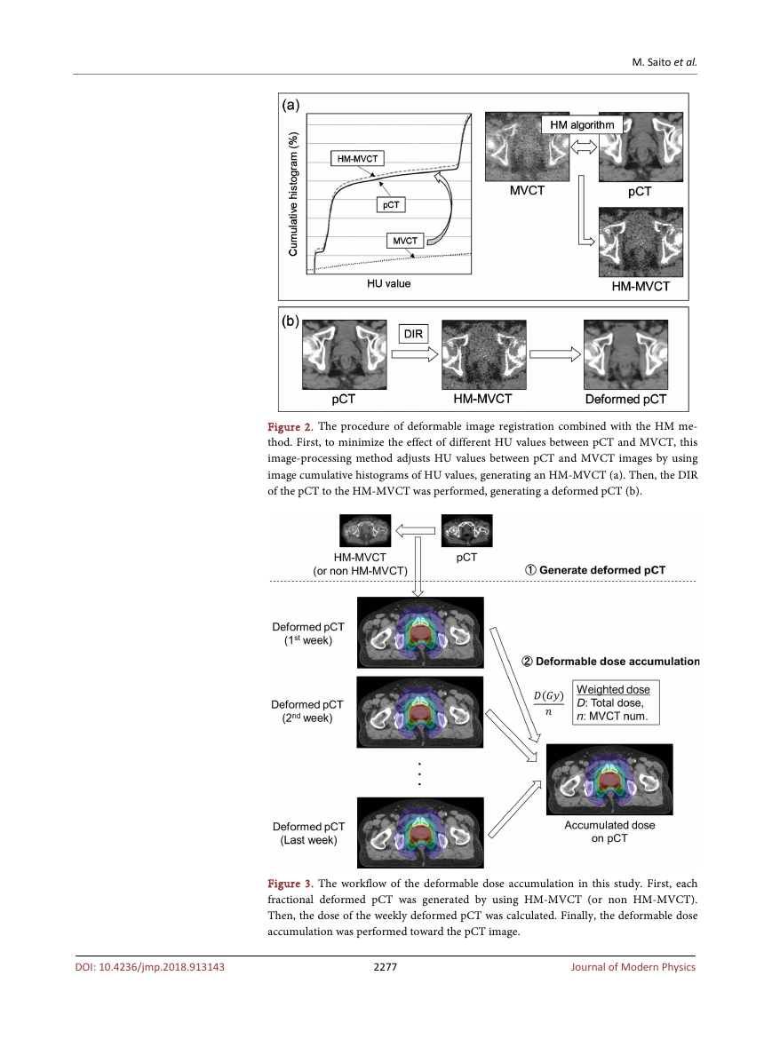
First, the DIR of pCT to HM-MVCT (or non-HM-MVCT) was performed. Fig-
ure 4 shows a typical case of improving the accuracy of deformation using HM
method for CTV. The accuracy of DIR was evaluated using DSC between pCT
Figure 4. A typical case of improving the accuracy of deformation using HM method for
CTV.
2278
Journal of Modern Physics
DOI: 10.4236/jmp.2018.913143
�
M. Saito et al.
and MVCT, comparing with the results of the non-HM method, as shown in
Figure 5. The mean DSC of the non-HM method was 0.75 ± 0.05, 0.83 ± 0.06,
and 0.90 ± 0.04 for the CTV, rectum, and bladder, respectively, while that of the
HM method was 0.81 ± 0.06, 0.81 ± 0.04, and 0.92 ± 0.06, respectively. The ac-
curacy for CTV and bladder was significantly improved compared with non-HM
MVCT (p < 0.05).
3.2. Dose Accumulation
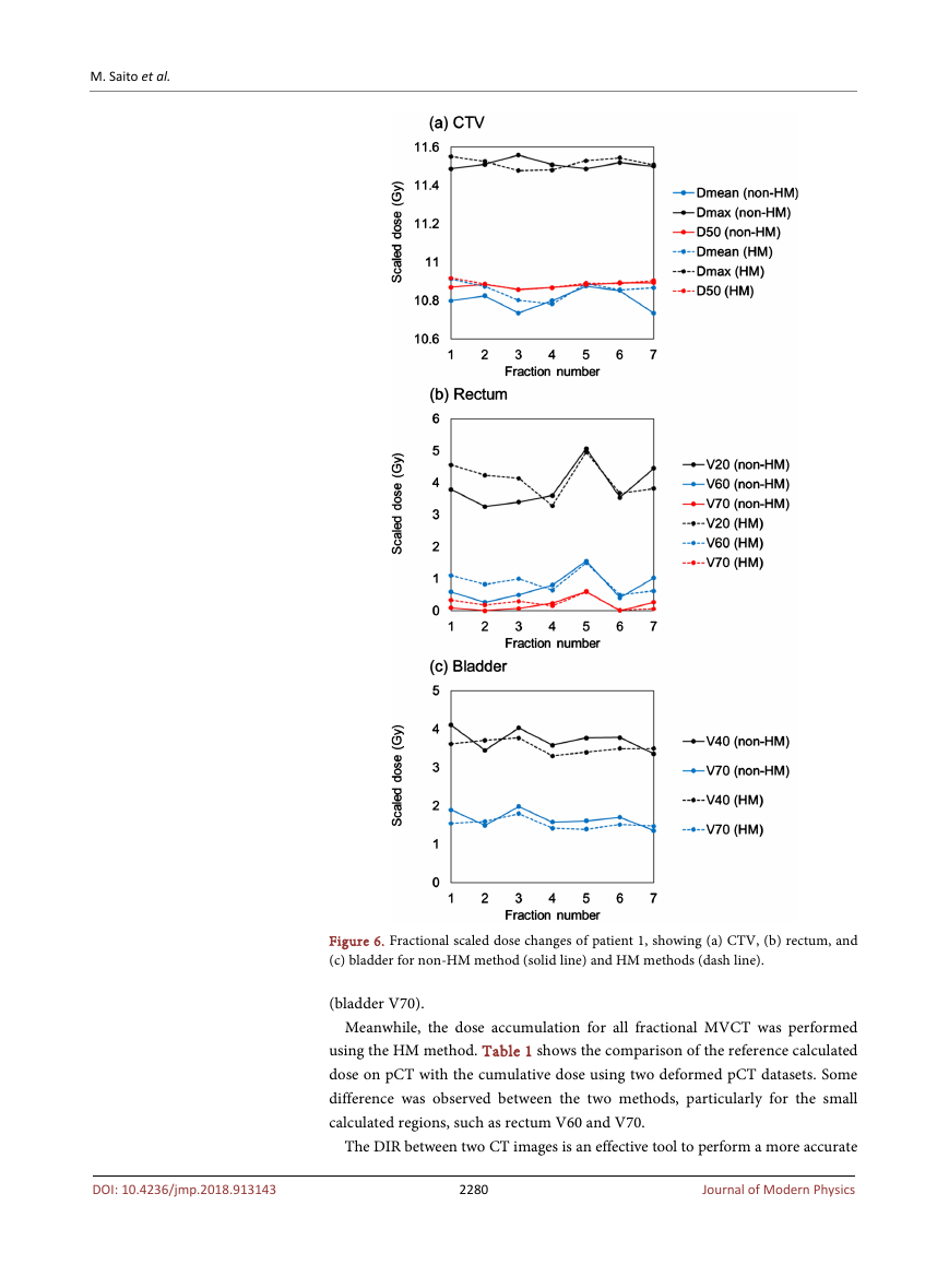
The dose calculation was performed for each fractional deformed pCT. Figure 6
shows the fractional scaled dose changes of patient 1, showing (a) CTV, (b) rec-
tum, and (c) bladder for non-HM method (solid line) and HM methods (dash
line). For all five patients, the maximum dose deviations of a same fraction be-
tween non-HM and HM methods were 0.16 Gy (CTV mean dose), −0.19 Gy
(CTV maximum dose), −0.16 Gy (CTV D50), −2.20 Gy (rectum V20), −1.62 Gy
(rectum V60), −1.23 Gy (rectum V70), 1.9 Gy (bladder V60), and 1.23 Gy
Figure 5. Patient individual dice similarity coefficient (DSC) between manual contours
on the each fractional MVCT and deformed contours on the deformed pCT for (a) CTV,
(b) rectum, and (c) bladder, and the average DSCs of all patients are shown in (d). Aste-
risk symbols (*) indicates a significant difference between two methods (p<0.05). The
mean DSC of the non-HM method was 0.75 ± 0.05, 0.83 ± 0.06, and 0.90 ± 0.04 for the
CTV, rectum, and bladder, respectively, while that of the HM method was 0.81 ± 0.06,
0.81 ± 0.04, and 0.92 ± 0.06, respectively.
2279
Journal of Modern Physics
DOI: 10.4236/jmp.2018.913143
�
M. Saito et al.
Figure 6. Fractional scaled dose changes of patient 1, showing (a) CTV, (b) rectum, and
(c) bladder for non-HM method (solid line) and HM methods (dash line).
(bladder V70).
Meanwhile, the dose accumulation for all fractional MVCT was performed
using the HM method. Table 1 shows the comparison of the reference calculated
dose on pCT with the cumulative dose using two deformed pCT datasets. Some
difference was observed between the two methods, particularly for the small
calculated regions, such as rectum V60 and V70.
The DIR between two CT images is an effective tool to perform a more accurate
2280
Journal of Modern Physics
DOI: 10.4236/jmp.2018.913143
�
Table 1. Comparison of the reference calculated dose on pCT with the cumulative dose
using two deformed pCT datasets. All p-values were calculated with Wilcoxon test.
M. Saito et al.
calculated accumulate dose (Gy, mean ± s.d.)
pCT
76.5 ± 0.1
79.7 ± 0.7
76.6 ± 0.1
41.1 ± 8.7
11.3 ± 5.4
5.0 ± 3.8
40.4 ± 14.0
15.2 ± 3.1
deformed pCT
(non-HM-method) p-value
0.71
0.08
1.00
0.26
0.01
0.01
0.17
0.01
76.4 ± 0.8
79.1 ± 0.8
76.6 ± 0.8
40.1 ± 8.1
7.1 ± 4.5
2.1 ± 3.2
38.5 ± 14.1
11.7 ± 3.3
deformed pCT
(HM-method) p-value
0.77
76.6 ± 0.9
0.37
79.3 ± 0.9
76.7 ± 0.9
0.70
0.08
43.1 ± 8.4
0.77
11.1 ± 5.0
0.34
4.3 ± 3.4
40.4 ± 15.7
0.99
0.47
15.7 ± 3.5
rectum
CTV Dmean
Dmax
D50
V20
V60
V70
V40
V70
bladder
adaptive radiotherapy. It is usually applied to two kV CT images (two pCT im-
ages or pCT image-daily CBCT image). Several authors evaluated their accuracy
by using DSC and target registration error (TRE) [11] [12] [13] [14] [15]. Al-
though various DIR algorithms were used for these validations, the accuracy of
DIR usually depends on the parameter setting of each software. In this study, we
used the intensity-based DIR implemented in MIM software. Kadoya et al. in-
vestigated the algorithm and found that the 3D registration error was 2.17 mm
to 3.61 mm in 10 theoretical images [14]. Although their results were not fo-
cused on the pelvic images, the accuracy of the deformation using MIM software
was comparable with that of the other commercial DIR software.
The MVCT is usually used for daily patient setup for image-guided HT treat-
ment instead of kV-CBCT implemented in a conventional linac. Some authors
investigated the use of daily MVCT to adaptive radiotherapy, for example, the
dose calculation and the deformation between kVCT to MVCT [5] [6]. Howev-
er, kVCT and MVCT have some differences, namely, imaging noise, contrast
resolution, and field of view, worsening the accuracy for intensity-based defor-
mation compared with kV-kV registration. Therefore, to improve the accuracy
of DIR, we applied HM algorithm between pCT and MVCT.
The HM algorithm is a method used to adjust HU values between pCT and
CBCT images by using image cumulative histograms. Van Zijtveld et al. showed
that the percentage of points with a gamma value < 1 (2%/2 mm) between pCT
and modified CBCT was 98% using the HM algorithm in five patients [10].
However, these reports focused on kV-CBCT, and reports on MVCT combined
with the HM algorithm have not been evaluated. Our results indicating that the
HM algorithm was effective in improving the accuracy of DIR between pCT and
MVCT for almost all anatomical structures are similar to that of the previous
kV-CBCT studies [8] [9] [13]. However, some cases had rectal gas and volume
change of the bladder, resulting in more variations in the accuracy of DIR. In
addition, one of the limitations of this study was the use of the merged MVCT
2281
Journal of Modern Physics
DOI: 10.4236/jmp.2018.913143
�




 2023年江西萍乡中考道德与法治真题及答案.doc
 2012年重庆南川中考生物真题及答案.doc
 2013年江西师范大学地理学综合及文艺理论基础考研真题.doc
 2020年四川甘孜小升初语文真题及答案I卷.doc
 2020年注册岩土工程师专业基础考试真题及答案.doc
 2023-2024学年福建省厦门市九年级上学期数学月考试题及答案.doc
 2021-2022学年辽宁省沈阳市大东区九年级上学期语文期末试题及答案.doc
 2022-2023学年北京东城区初三第一学期物理期末试卷及答案.doc
 2018上半年江西教师资格初中地理学科知识与教学能力真题及答案.doc
 2012年河北国家公务员申论考试真题及答案-省级.doc
 2020-2021学年江苏省扬州市江都区邵樊片九年级上学期数学第一次质量检测试题及答案.doc
 2022下半年黑龙江教师资格证中学综合素质真题及答案.doc