2010 Second International conference on Computing, Communication and Networking Technologies
A ROBUST FUZZY CLUSTERING TECHNIQUE WITH SPATIAL NEIGHBORHOOD
INFORMATION FOR EFFECTIVE MEDICAL IMAGE SEGMENTATION
An Efficient Variants of Fuzzy Clustering Technique with Spatial Information
for Effective Noisy Medical Image Segmentation
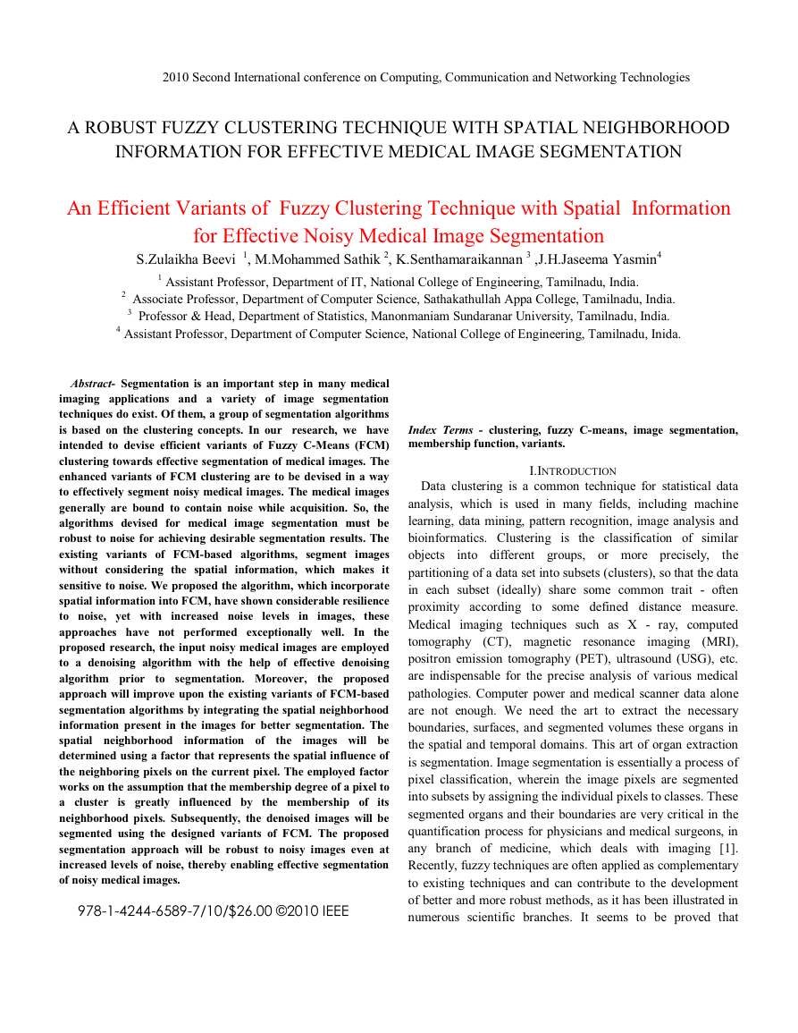
S.Zulaikha Beevi 1, M.Mohammed Sathik 2, K.Senthamaraikannan 3 ,J.H.Jaseema Yasmin4
1 Assistant Professor, Department of IT, National College of Engineering, Tamilnadu, India.
2 Associate Professor, Department of Computer Science, Sathakathullah Appa College, Tamilnadu, India.
3 Professor & Head, Department of Statistics, Manonmaniam Sundaranar University, Tamilnadu, India.
4 Assistant Professor, Department of Computer Science, National College of Engineering, Tamilnadu, Inida.
Abstract- Segmentation is an important step in many medical
imaging applications and a variety of image segmentation
techniques do exist. Of them, a group of segmentation algorithms
is based on the clustering concepts. In our research, we have
intended to devise efficient variants of Fuzzy C-Means (FCM)
clustering towards effective segmentation of medical images. The
enhanced variants of FCM clustering are to be devised in a way
to effectively segment noisy medical images. The medical images
generally are bound to contain noise while acquisition. So, the
algorithms devised for medical image segmentation must be
robust to noise for achieving desirable segmentation results. The
existing variants of FCM-based algorithms, segment images
without considering the spatial information, which makes it
sensitive to noise. We proposed the algorithm, which incorporate
spatial information into FCM, have shown considerable resilience
to noise, yet with increased noise levels in images, these
approaches have not performed exceptionally well. In the
proposed research, the input noisy medical images are employed
to a denoising algorithm with the help of effective denoising
algorithm prior to segmentation. Moreover, the proposed
approach will improve upon the existing variants of FCM-based
segmentation algorithms by integrating the spatial neighborhood
information present in the images for better segmentation. The
spatial neighborhood
images will be
determined using a factor that represents the spatial influence of
the neighboring pixels on the current pixel. The employed factor
works on the assumption that the membership degree of a pixel to
a cluster is greatly influenced by the membership of its
neighborhood pixels. Subsequently, the denoised images will be
segmented using the designed variants of FCM. The proposed
segmentation approach will be robust to noisy images even at
increased levels of noise, thereby enabling effective segmentation
of noisy medical images.
information of
the
978-1-4244-6589-7/10/$26.00 ©2010 IEEE
Index Terms - clustering, fuzzy C-means, image segmentation,
membership function, variants.
I.INTRODUCTION
into different groups, or more precisely,
Data clustering is a common technique for statistical data
analysis, which is used in many fields, including machine
learning, data mining, pattern recognition, image analysis and
bioinformatics. Clustering is the classification of similar
objects
the
partitioning of a data set into subsets (clusters), so that the data
in each subset (ideally) share some common trait - often
proximity according to some defined distance measure.
Medical imaging techniques such as X - ray, computed
tomography (CT), magnetic resonance
imaging (MRI),
positron emission tomography (PET), ultrasound (USG), etc.
are indispensable for the precise analysis of various medical
pathologies. Computer power and medical scanner data alone
are not enough. We need the art to extract the necessary
boundaries, surfaces, and segmented volumes these organs in
the spatial and temporal domains. This art of organ extraction
is segmentation. Image segmentation is essentially a process of
pixel classification, wherein the image pixels are segmented
into subsets by assigning the individual pixels to classes. These
segmented organs and their boundaries are very critical in the
quantification process for physicians and medical surgeons, in
any branch of medicine, which deals with imaging [1].
Recently, fuzzy techniques are often applied as complementary
to existing techniques and can contribute to the development
of better and more robust methods, as it has been illustrated in
numerous scientific branches. It seems to be proved that
�
applications of fuzzy techniques are very successful in the area
of image processing [2]. Moreover, the field of medicine has
become a very attractive domain for the application of fuzzy
set theory. This is due to the large role imprecision and
uncertainty plays in the field [3]. The main objective of
medical image segmentation is to extract and characterize
anatomical structures with respect to some input features or
expert knowledge. Segmentation methods that includes the
classification of tissues in medical imagery can be performed
using a variety of techniques. Many clustering strategies have
been used, such as the crisp clustering scheme and the fuzzy
clustering scheme, each of which has its own special
characteristics [3]. The conventional crisp clustering method
restricts each point of the data set to exclusively just one
cluster. However, in many real situations, for images, issues
such as limited spatial resolution, poor contrast, overlapping
intensities, noise and intensity in homogeneities variation
make this hard (crisp) segmentation a difficult task. The fuzzy
set theory [4], which involves the idea of partial membership
described by a membership function, fuzzy clustering as a soft
segmentation method has been widely studied and successfully
applied to image segmentation [5–11]. Among the fuzzy
clustering methods, fuzzy c-means (FCM) algorithm [1] is the
most popular method used in image segmentation because it
has robust characteristics for ambiguity and can retain much
more
than hard segmentation methods [5,
6].Although the conventional FCM algorithm works well on
most noise-free images, it has a serious limitation: it does not
incorporate any information about spatial context, which cause
it to be sensitive to noise and imaging artifacts. In this paper,
Improved Spatial FCM (ISFCM) clustering algorithm for
image segmentation is proposed. The algorithm is developed
by incorporating the spatial neighborhood information into the
standard FCM clustering algorithm by a priori probability.
The probability is given to indicate the spatial influence of the
neighboring pixels on
the centre pixel, which can be
automatically decided in the implementation of the algorithm
by the fuzzy membership. The new fuzzy membership of the
current centre pixel is then recalculated with this probability
obtained from above. The algorithm is initialized by a given
histogram based FCM algorithm, which helps to speed up the
convergence of the algorithm. Experimental results with
medical images that the proposed method can achieve
comparable results to those from many derivatives of FCM
algorithm, that gives the method presented in this paper is
effective.
information
II. RELATED WORKS
There are huge amount of works related to enhancing the
conventional FCM and other forms for image segmentation are
found in the literature. Let us review some of them. Smaine
Mazouzi and Mohamed Batouche [13] have presented an
approach for improving range image segmentation, based on
fuzzy regularization of the detected edges. Initially, a degraded
version of the segmentation was produced by a new region
growing- based algorithm. Next, the resulting segmentation
was refined by a robust fuzzy classification of the pixels on the
resulting edges which correspond to border of the extracted
regions. Pixels on the boundary between two adjacent regions
are labeled taking into account the two regions as fuzzy sets in
the fuzzy classification stage, using an improved version of the
Fuzzy C-Mean algorithm. The process was repeated for all
region boundaries in the image. A two-dimensional fuzzy C-
means (2DFCM) algorithm was proposed by Jinhua Yu and
Yuanyuan Wang [14] for the molecular image segmentation.
The 2DFCM algorithm was composed of three stages. The first
stage was the noise suppression by utilizing a method
combining a Gaussian noise filter and anisotropic diffusion
techniques. The second stage was
texture energy
characterization using a Gabor wavelet method. The third
stage was introducing spatial constraints provided by the
denoising data and the textural information into the two-
dimensional fuzzy clustering. The incorporation of intensity
and textural information allows the 2DFCM algorithm to
produce satisfactory segmentation results for images corrupted
by noise (outliers) and intensity variations. Hadi Sadoghi
Yazdi and Jalal A. Nasiri [15] have presented a fuzzy image
segmentation algorithm. In their algorithm, human knowledge
was used in clustering features for fuzzy image segmentation.
In fuzzy clustering, the membership values of extracted
features for each pixel at each cluster change proportional to
zonal mean of membership values and gradient mean of
adjacent pixels. The direction of membership variations are
interaction. Their segmentation
specified using human
approach was applied for segmentation of
texture and
documentation images and the results have shown that the
human interaction eventuates to clarification of texture and
reduction of noise in segmented images.G.Sudhavani and
Dr.K.Sathyaprasad [16] have described the application of a
modified fuzzy C-means clustering algorithm to the lip
segmentation problem. The modified fuzzy C-means algorithm
was able to take the initial membership function from the
spatially
Successful
segmentation of lip images was possible with their method.
Comparative study of their modified fuzzy C-means was done
with the traditional fuzzy C-means algorithm by using Pratt’s
Figure of Merit. (2009) B.Sowmya and B.Sheelarani [9] have
explained the task of segmenting any given color image using
soft computing techniques. The most basic attribute for
segmentation was
for a
luminance amplitude
neighboring
image
the
connected
pixels.
�
the need
monochrome image and color components for a color image.
Since there are more than 16 million colors available in any
given image and it was difficult to analyze the image on all of
its colors, the likely colors are grouped together by image
segmentation. For that purpose soft computing techniques have
been used. The soft computing techniques used are Fuzzy C-
Means algorithm (FCM), Possibilistic C - Means algorithm
(PCM) and competitive neural network. A self estimation
algorithm was developed for determining the number of
clusters. Agus Zainal Arifin and Akira Asano [12] have
proposed a method of image thresholding by using cluster
organization from the histogram of an image. A similarity
measure proposed by them was based on inter-class variance
of the clusters to be merged and the intra-class variance of the
new merged cluster. Experiments on practical images have
illustrated the effectiveness of their method. (2006) An high
speed parallel fuzzy C means algorithm for brain tumor image
segmentation is presented by S. Murugavalli and V. Rajamani
[17]. Their algorithm has the advantages of both the sequential
FCM and parallel FCM for the clustering process in the
segmentation techniques and the algorithm was very fast when
the image size was large and also it requires less execution
time. They have also achieved less processing speed and
minimizing
for accessing secondary storage
compared to the previous results. The reduction in the
computation time was primarily due to the selection of actual
cluster centre and the accessing minimum secondary storage.
(2006) T. Bala Ganesan and R. Sukanesh [18] have deals with
Brain Magnetic Resonance Image Segmentation. Any medical
image of human being consists of distinct regions and these
regions could be
represented by wavelet coefficients.
Classification of these features was performed using Fuzzy
Clustering method (FCM Fuzzy C-Means Algorithm). Edge
detection technique was used to detect the edges of the given
images. Silhouette method was used to find the strength of
clusters. Finally, the different regions of the images are
demarcated and color coded. (2008) H. C. Sateesh Kumar et al.
[19] have proposed Automatic Image Segmentation using
Wavelets (AISWT) to make segmentation fast and simpler.
The approximation band of image Discrete Wavelet Transform
was considered for segmentation which contains significant
information of
image. The Histogram based
algorithm was used to obtain the number of regions and the
initial parameters like mean, variance and mixing factor. The
final parameters are obtained by using the Expectation and
Maximization
the
approximation coefficients was determined by Maximum
Likelihood function. Histogram specification was proposed by
Gabriel Thomas [20] as a way to improve image segmentation.
Specification of the final histogram was done relatively easy
algorithm. The
segmentation
the
input
of
and all it takes is the definition of a low pass filter and the
amplification and attenuation of the peaks and valleys
respectively or the standard deviation of the assumed Gaussian
modes in the final specification. Examples showing better
segmentation were presented. The attractive side of their
approach was the easy implementation that was needed to
obtain considerable better results during the segmentation
process.
III. DESCRIPTION OF EMPLOYED DENOISING ALGORITHM
Usually, the medical images obtained from sensors are bound
to contain noise and blurred edges. The process of
segmentation is made more intricate, owing to the presence of
these artifacts in medical images. Consequently, denoising
images prior
to segmentation perhaps produce better
segmentation accuracy. Recently, Alessandro Foi et al [21]
presented an efficient denoising algorithm, which is used in the
proposed approach. Initially, the input noisy medical images
are denoised using the above-mentioned denoising algorithm.
A brief description of the denoising strategy employed in the
proposed approach is provided in the subsequent subsection.
A. Pointwise SA-DCT Denoising
Since noise is an inevitable one in image acquisition,
denoising plays an important role in increasing the quality of
the image. Noise removal has been widely studied as a
primary low-level image processing procedure and copious
amount of denoising schemes have been proposed. In our
approach, we employed an efficient Pointwise Shape Adaptive
DCT denoising algorithm[21] . In order to preserve the image
local structures in a better way, within the transform support.
In this way, it ensures that data are represented sparsely in the
transform domain, significantly improving the effectiveness of
thresholding. Before we proceed
is worth
mentioning that the approach can be interpreted as a special
kind of local model selection which is adaptive with respect
both to the scale and to the order of the utilized model. Shape-
adapted orthogonal polynomials are the most obvious choice
for the local transform, as they are more consistent with the
polynomial modeling used to determine its support. However,
in practice, cosine bases are known to be more adequate for the
modeling of natural images. In particular, when image
processing applications are of concern,
the use of
computationally efficient transforms is paramount and, thus,
the low-complexity SA-DCT and high robustness to noise.
further,
it
IV. PROPOSED APPROACH FOR SPATIAL FUZZY C-MEANS
CLUSTERING
�
A. The Conventional FCM
Clustering is the process of finding groups in unlabeled
dataset based on a similarity measure between the data patterns
(elements) [12]. A cluster contains similar patterns placed
together. The fuzzy clustering technique generates fuzzy
partitions of the data instead of hard partitions. Therefore, data
patterns may belong to several clusters, having different
membership values with different clusters. The membership
value of a data pattern to a cluster denotes similarity between
the given data pattern to the cluster. Given a set of n data
patterns, X = x1,…,xk,…,xn, the fuzzy clustering technique
minimizes the objective function, O(U,C ):
fcmO
)C(U,
=
(
)
v
n
m
)ic,k(x2d
∑
∑
iku
=
=
i
k
1 1
(1)
where xk is the k-th D-dimensional data vector, ci is the
center of cluster i, uik is the degree of membership of xk in the
i-th cluster, m is the weighting exponent, d (xk, ci) is the
distance between data xk and cluster center ci, n is the number
of data patterns, v is the number of clusters. The minimization
of objective function J (U, C) can be brought by an iterative
process in which updating of degree of membership uik and the
cluster centers are done for each iteration.
in
information
The theory of Markov random field says that pixels in the
image mostly belong to the same cluster as their neighbors.
The incorporation of spatial information in the clustering
process makes the algorithm robust to noise and blurred edges.
But when using spatial
the clustering
optimization function may converge in local minima, so to
avoid this problem the fuzzy spatial c means algorithm is
initialized with the Histogram based fuzzy c-means algorithm.
The optimization function for
histogram based fuzzy
clustering is given in the equation 4.
(
L
v
∑
∑
ilu
=
=
i
1 1
)
)ic,(2d)H(m
(4)
hfcmO
)C
(U,
l
=
l
where H is the histogram of the image of L-gray levels.
Gray level of all the pixels in the image lies in the new discrete
set G= {0,1,…,L-1}. The computation of membership degrees
of H(l) pixels is reduced to that of only one pixel with l as grey
ilu and center for
level value. The member ship function
histogram based fuzzy c-means clustering can be calculated as.
l
=
ilu
=
iku
1
⎛V
⎜
∑
⎜
=
J
1
⎝
ikd
jkd
⎞
⎟
⎟
⎠
1
−
m
1
ic
L
∑
==
l
1
1
⎛V
⎜
∑
⎜
=
J
1
⎝
lid
ljd
(
)
m
ilu
(
L
∑
ilu
=
l
1
(5)
1
−
m
1
⎞
⎟
⎟
⎠
l
( )
lH
)
m
(2)
(
n
∑
iku
==
k
1
(
n
∑
iku
=
k
1
)
m
kx
)
m
ic
(3)
i∀ iku satisfies:
]1,0∈iku
[
,
v
k∀ ∑
iku
=
i
1
=
1 and
iku
<
n
where
<
0
n
∑
=
k
1
Thus the conventional clustering technique clusters an
image data only with the intensity values but it does not use
the spatial information of the given image.
B. Initialization
(6)
where
level l
lid is the distance between the center i and the gray
C. Proposed Approach
As histogram based FCM algorithm operates merely on the
histogram of an image, it is faster than the conventional FCM
that processes the entire data [22]. Despite the fact that,
conventional FCM algorithm works well on the majority of
noise-free images, it has a major drawback, (i.e.) it is highly
sensitive to noise and many other imaging artifacts. The
histogram-based FCM can be made more robust against noise
and blurred edges by incorporating the spatial information into
it. The objective
the proposed
segmentation approach is given by
)CO(U,
function
of
�
fcmO
)C(U,
=
(
v
n
s
∑
∑
iku
=
=
i
k
1 1
)
m
)ic,k(x2d
(7)
s
iku
=
1
−
m
1
V
∑
=
J
1
⎛
⎜
⎜
⎝
ikd
jkd
⎞
⎟
⎟
⎠
⎛
⎜
⎜
⎜
⎜
⎝
ikP
kN
∑
=
z
1
kN
⎛
⎜
⎜
⎜⎜
⎝
⎛
⎞
⎜
⎟
⎜
⎟
⎜
⎟
⎜
⎟
⎝
⎠
1
−
m
1
V
∑
=
J
1
⎛
⎜
⎜
⎝
izd
jzd
⎞
⎟
⎟
⎠
The spatial membership function
is calculated using the equation (8).
iku of the proposed ISFCM
s
ikP is the apriori probability that kth pixel belongs to ith
where
cluster and calculated as
ikP =
( )
kiNN
kN
(9)
where NNi(k) is the number of pixels in the neighborhood of
kN is
kth pixel that belongs to cluster i after defuzzification.
izd is the
the total number of pixels in the neighborhood.
distance between ith cluster and zth neighborhood of ith Thus the
center
ic of each cluster is calculated as
s
s
ic
(
)
n
ms
∑
iku
kx
==
k
1
(
)
n
ms
∑
iku
=
k
1
(10)
Two kinds of spatial information are incorporated in the
member ship
function of conventional FCM. Apriori
probability and Fuzzy spatial information
Apriori probability: This parameter assigns a noise pixel to
one of the clusters to which its neighborhood pixels belong.
The noise pixel is included in the cluster whose members are
majority in the pixels neighborhood.
Fuzzy spatial information: In the equation (8) the second term
in the denominator is the average of fuzzy membership of the
neighborhood pixel to a cluster. Thus a pixel gets higher
membership value when their neighborhood pixels have high
membership value with the corresponding cluster.
V. RESULTS AND DISCUSSION
The proposed algorithm converges very quickly because it
gets initial parameters form already converged histogram
⎞
⎞
⎟
⎟
⎟
⎟
⎟
⎟⎟
⎟
⎠
⎠
(8)
based FCM. The proposed approach is applied on three kinds
of images real world images, synthetic brain MRI image,
original brain MRI image. In all the images additive Gaussian
white noise is added with noise percentage level 0%, 5%, 10%,
and 15% and corresponding results are shown. The quality of
segmentation of the proposed algorithm can be calculated by
segmentation accuracy As given as.
=
sA
100×
cN
pT
(11)
Nc is the number of correctly classified pixels and Tp is the
total
image.
Segmentation accuracy of FCM, proposed approach without
denoising in segmenting synthetic brain MRI images with
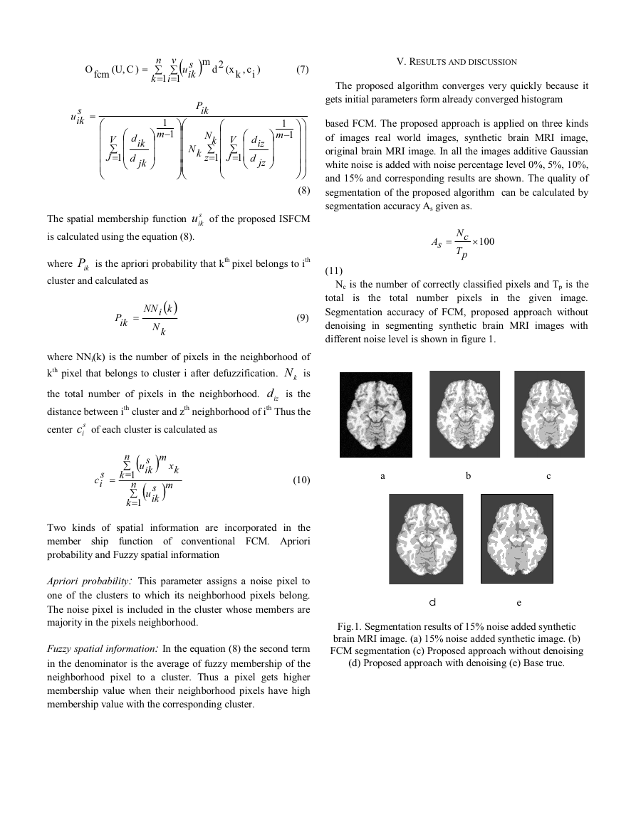
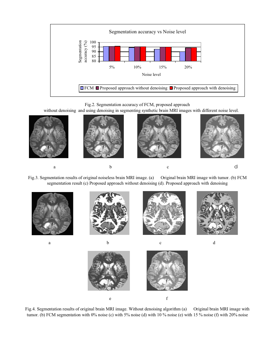
different noise level is shown in figure 1.
total number pixels
the given
is
the
in
a b c
d e
Fig.1. Segmentation results of 15% noise added synthetic
brain MRI image. (a) 15% noise added synthetic image. (b)
FCM segmentation (c) Proposed approach without denoising
(d) Proposed approach with denoising (e) Base true.
�
)
%
n
o
i
t
a
t
n
e
m
g
e
S
(
y
c
a
r
u
c
c
a
100
95
90
85
80
Segmentation accuracy vs Noise level
5%
10%
15%
20%
Noise level
FCM Proposed approach without denoising
Proposed approach with denoising
without denoising and using denoising in segmenting synthetic brain MRI images with different noise level.
Fig.2. Segmentation accuracy of FCM, proposed approach
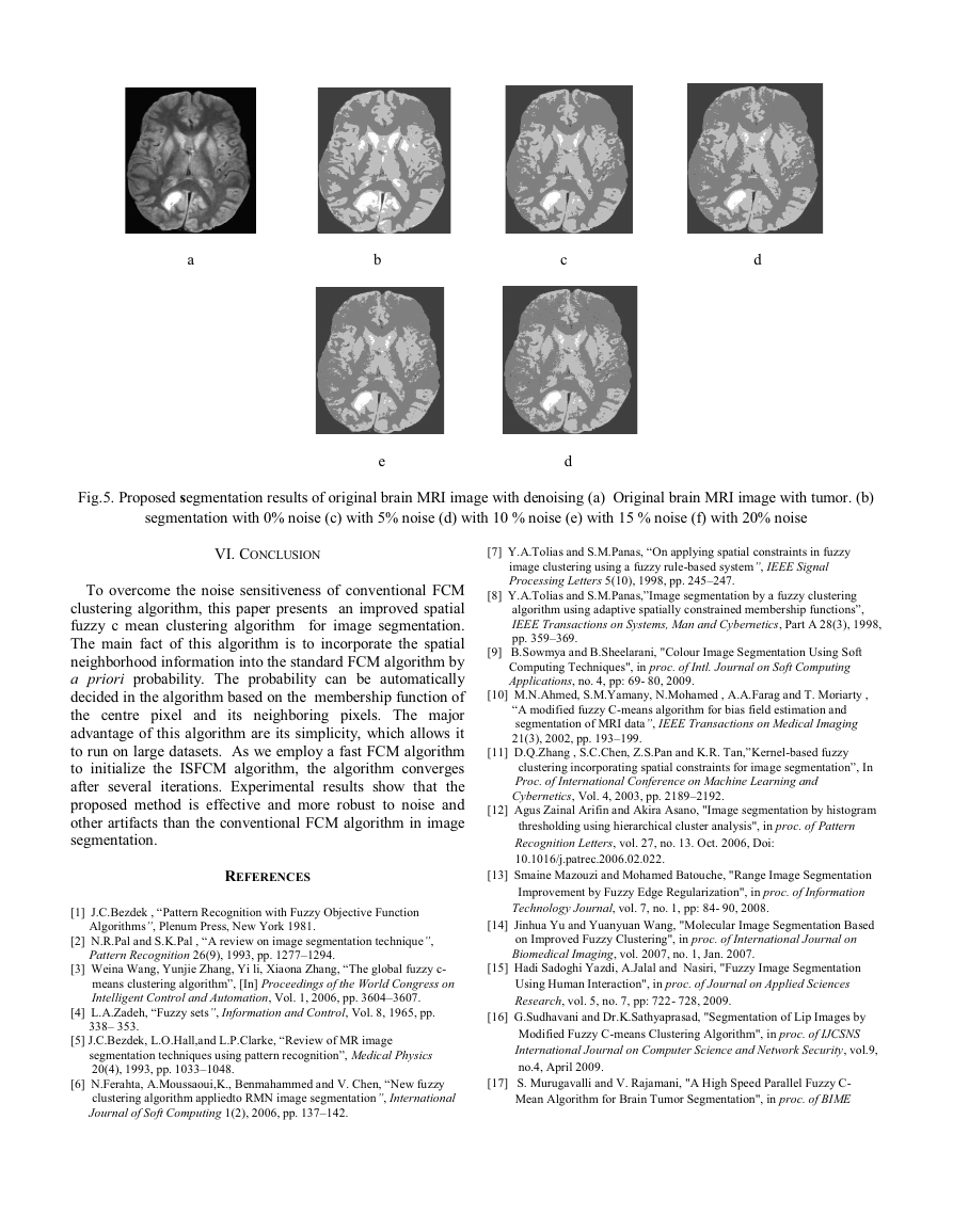
a b c d
Fig.3. Segmentation results of original noiseless brain MRI image. (a) Original brain MRI image with tumor. (b) FCM
segmentation result (c) Proposed approach without denoising (d). Proposed approach with denoising
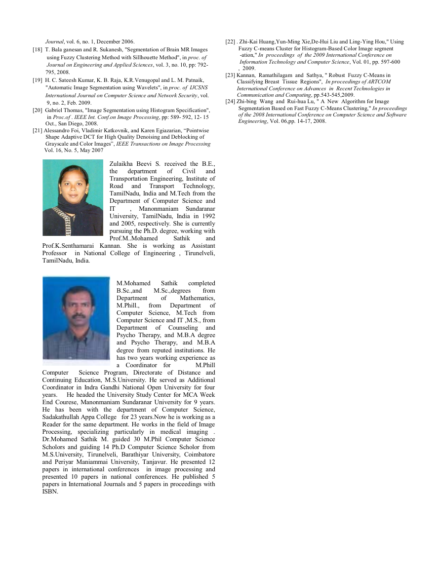
a b c d
e f
Fig.4. Segmentation results of original brain MRI image. Without denoising algorithm (a) Original brain MRI image with
tumor. (b) FCM segmentation with 0% noise (c) with 5% noise (d) with 10 % noise (e) with 15 % noise (f) with 20% noise
�
a b c d
e d
Fig.5. Proposed segmentation results of original brain MRI image with denoising (a) Original brain MRI image with tumor. (b)
segmentation with 0% noise (c) with 5% noise (d) with 10 % noise (e) with 15 % noise (f) with 20% noise
VI. CONCLUSION
To overcome the noise sensitiveness of conventional FCM
clustering algorithm, this paper presents an improved spatial
fuzzy c mean clustering algorithm for image segmentation.
The main fact of this algorithm is to incorporate the spatial
neighborhood information into the standard FCM algorithm by
a priori probability. The probability can be automatically
decided in the algorithm based on the membership function of
the centre pixel and its neighboring pixels. The major
advantage of this algorithm are its simplicity, which allows it
to run on large datasets. As we employ a fast FCM algorithm
to initialize the ISFCM algorithm, the algorithm converges
after several iterations. Experimental results show that the
proposed method is effective and more robust to noise and
other artifacts than the conventional FCM algorithm in image
segmentation.
REFERENCES
[1] J.C.Bezdek , “Pattern Recognition with Fuzzy Objective Function
Algorithms”, Plenum Press, New York 1981.
[2] N.R.Pal and S.K.Pal , “A review on image segmentation technique”,
Pattern Recognition 26(9), 1993, pp. 1277–1294.
[3] Weina Wang, Yunjie Zhang, Yi li, Xiaona Zhang, “The global fuzzy c-
means clustering algorithm”, [In] Proceedings of the World Congress on
Intelligent Control and Automation, Vol. 1, 2006, pp. 3604–3607.
[4] L.A.Zadeh, “Fuzzy sets”, Information and Control, Vol. 8, 1965, pp.
338– 353.
[5] J.C.Bezdek, L.O.Hall,and L.P.Clarke, “Review of MR image
segmentation techniques using pattern recognition”, Medical Physics
20(4), 1993, pp. 1033–1048.
[6] N.Ferahta, A.Moussaoui,K., Benmahammed and V. Chen, “New fuzzy
clustering algorithm appliedto RMN image segmentation”, International
Journal of Soft Computing 1(2), 2006, pp. 137–142.
[7] Y.A.Tolias and S.M.Panas, “On applying spatial constraints in fuzzy
image clustering using a fuzzy rule-based system”, IEEE Signal
Processing Letters 5(10), 1998, pp. 245–247.
[8] Y.A.Tolias and S.M.Panas,”Image segmentation by a fuzzy clustering
algorithm using adaptive spatially constrained membership functions”,
IEEE Transactions on Systems, Man and Cybernetics, Part A 28(3), 1998,
pp. 359–369.
[9] B.Sowmya and B.Sheelarani, "Colour Image Segmentation Using Soft
Computing Techniques", in proc. of Intl. Journal on Soft Computing
Applications, no. 4, pp: 69- 80, 2009.
[10] M.N.Ahmed, S.M.Yamany, N.Mohamed , A.A.Farag and T. Moriarty ,
“A modified fuzzy C-means algorithm for bias field estimation and
segmentation of MRI data”, IEEE Transactions on Medical Imaging
21(3), 2002, pp. 193–199.
[11] D.Q.Zhang , S.C.Chen, Z.S.Pan and K.R. Tan,”Kernel-based fuzzy
clustering incorporating spatial constraints for image segmentation”, In
Proc. of International Conference on Machine Learning and
Cybernetics, Vol. 4, 2003, pp. 2189–2192.
[12] Agus Zainal Arifin and Akira Asano, "Image segmentation by histogram
thresholding using hierarchical cluster analysis", in proc. of Pattern
Recognition Letters, vol. 27, no. 13. Oct. 2006, Doi:
10.1016/j.patrec.2006.02.022.
[13] Smaine Mazouzi and Mohamed Batouche, "Range Image Segmentation
Improvement by Fuzzy Edge Regularization", in proc. of Information
Technology Journal, vol. 7, no. 1, pp: 84- 90, 2008.
[14] Jinhua Yu and Yuanyuan Wang, "Molecular Image Segmentation Based
on Improved Fuzzy Clustering", in proc. of International Journal on
Biomedical Imaging, vol. 2007, no. 1, Jan. 2007.
[15] Hadi Sadoghi Yazdi, A.Jalal and Nasiri, "Fuzzy Image Segmentation
Using Human Interaction", in proc. of Journal on Applied Sciences
Research, vol. 5, no. 7, pp: 722- 728, 2009.
[16] G.Sudhavani and Dr.K.Sathyaprasad, "Segmentation of Lip Images by
Modified Fuzzy C-means Clustering Algorithm", in proc. of IJCSNS
International Journal on Computer Science and Network Security, vol.9,
no.4, April 2009.
[17] S. Murugavalli and V. Rajamani, "A High Speed Parallel Fuzzy C-
Mean Algorithm for Brain Tumor Segmentation", in proc. of BIME
�
Journal, vol. 6, no. 1, December 2006.
[18] T. Bala ganesan and R. Sukanesh, "Segmentation of Brain MR Images
using Fuzzy Clustering Method with Sillhouette Method", in proc. of
Journal on Engineering and Applied Sciences, vol. 3, no. 10, pp: 792-
795, 2008.
[19] H. C. Sateesh Kumar, K. B. Raja, K.R.Venugopal and L. M. Patnaik,
"Automatic Image Segmentation using Wavelets", in proc. of IJCSNS
International Journal on Computer Science and Network Security, vol.
9, no. 2, Feb. 2009.
[20] Gabriel Thomas, "Image Segmentation using Histogram Specification",
in Proc.of . IEEE Int. Conf.on Image Processing, pp: 589- 592, 12- 15
Oct., San Diego, 2008.
[21] Alessandro Foi, Vladimir Katkovnik, and Karen Egiazarian, “Pointwise
Shape Adaptive DCT for High Quality Denoising and Deblocking of
Grayscale and Color Images”, IEEE Transactions on Image Processing
Vol. 16, No. 5, May 2007
[22] . Zhi-Kai Huang,Yun-Ming Xie,De-Hui Liu and Ling-Ying Hou," Using
Fuzzy C-means Cluster for Histogram-Based Color Image segment
-ation," In proceedings of the 2009 International Conference on
Information Technology and Computer Science, Vol. 01, pp. 597-600
, 2009.
[23] Kannan, Ramathilagam and Sathya, " Robust Fuzzy C-Means in
Classifying Breast Tissue Regions", In proceedings of ARTCOM
International Conference on Advances in Recent Technologies in
Communication and Computing, pp.543-545,2009.
[24] Zhi-bing Wang and Rui-hua Lu, " A New Algorithm for Image
Segmentation Based on Fast Fuzzy C-Means Clustering," In proceedings
of the 2008 International Conference on Computer Science and Software
Engineering, Vol. 06,pp. 14-17, 2008.
of Civil
department
Zulaikha Beevi S. received the B.E.,
the
and
Transportation Engineering, Institute of
Road
and Transport Technology,
TamilNadu, India and M.Tech from the
Department of Computer Science and
IT
, Manonmaniam Sundaranar
University, TamilNadu, India in 1992
and 2005, respectively. She is currently
pursuing the Ph.D. degree, working with
Prof.M..Mohamed
and
Prof.K.Senthamarai Kannan. She
is working as Assistant
Professor in National College of Engineering , Tirunelveli,
TamilNadu, India.
Sathik
of
for
Sathik
from Department
completed
M.Mohamed
from
B.Sc.,and M.Sc.,degrees
Mathematics,
Department
M.Phill.,
of
Computer Science, M.Tech from
Computer Science and IT ,M.S., from
Department of Counseling
and
Psycho Therapy, and M.B.A degree
and Psycho Therapy, and M.B.A
degree from reputed institutions. He
has two years working experience as
a Coordinator
M.Phill
Computer
Science Program, Directorate of Distance and
Continuing Education, M.S.University. He served as Additional
Coordinator in Indra Gandhi National Open University for four
years. He headed the University Study Center for MCA Week
End Courese, Manonmaniam Sundaranar University for 9 years.
He has been with the department of Computer Science,
Sadakathullah Appa College for 23 years.Now he is working as a
Reader for the same department. He works in the field of Image
Processing, specializing particularly
.
Dr.Mohamed Sathik M. guided 30 M.Phil Computer Science
Scholors and guiding 14 Ph.D Computer Science Scholor from
M.S.University, Tirunelveli, Barathiyar University, Coimbatore
and Periyar Maniammai University, Tanjavur. He presented 12
papers in international conferences in image processing and
presented 10 papers in national conferences. He published 5
papers in International Journals and 5 papers in proceedings with
ISBN.
in medical
imaging
�


 2023年江西萍乡中考道德与法治真题及答案.doc
 2012年重庆南川中考生物真题及答案.doc
 2013年江西师范大学地理学综合及文艺理论基础考研真题.doc
 2020年四川甘孜小升初语文真题及答案I卷.doc
 2020年注册岩土工程师专业基础考试真题及答案.doc
 2023-2024学年福建省厦门市九年级上学期数学月考试题及答案.doc
 2021-2022学年辽宁省沈阳市大东区九年级上学期语文期末试题及答案.doc
 2022-2023学年北京东城区初三第一学期物理期末试卷及答案.doc
 2018上半年江西教师资格初中地理学科知识与教学能力真题及答案.doc
 2012年河北国家公务员申论考试真题及答案-省级.doc
 2020-2021学年江苏省扬州市江都区邵樊片九年级上学期数学第一次质量检测试题及答案.doc
 2022下半年黑龙江教师资格证中学综合素质真题及答案.doc