CiA Draft Standard 412
CANopen
Profiles for medical devices
Part 2: Automatic X-ray collimator
Version 1.0
31 December 2005
© CAN in Automation (CiA) e. V.
�
DS 412-2 V1.0 CANopen profiles for medical devices - Automatic X-ray collimator
HISTORY
CiA
Date
Changes
25.05. 2003
31.12. 2005
Publication of Version 1.0 as Draft Standard Proposal
Publication of Version 1.0 as Draft Standard
General information on licensing and patents
CAN in AUTOMATION (CiA) calls attention to the possibility that some of the elements of this CiA
specification may be subject of patent rights. CiA shall not be responsible for identifying any or all such
patent rights.
Because this specification is licensed free of charge, there is no warranty for this
specification, to the extent permitted by applicable law. Except when otherwise stated in
writing the copyright holder and/or other parties provide this specification “as is” without
warranty of any kind, either expressed or implied, including, but not limited to, the implied
warranties of merchantability and fitness for a particular purpose. The entire risk as to the
correctness and completeness of the specification is with you. Should this specification prove
failures, you assume the cost of all necessary servicing, repair or correction.
© CiA 2008
All rights reserved. Unless otherwise specified, no part of this publication may be reproduced or
utilized in any form or by any means, electronic or mechanical, including photocopying and microfilm,
without permission in writing from CiA at the address below.
CAN in Automation e. V.
Kontumazgarten 3
DE - 90429 Nuremberg, Germany
Tel.: +49-911-928819-0
Fax: +49-911-928819-79
Url: www.can-cia.org
Email: headquarters@can-cia.org
2
© CiA 2008 – All rights reserved
�
DS 412-2 V1.0 CANopen profiles for medical devices - Automatic X-ray collimator
CiA
CONTENTS
5.1
5.2
5.2.1
5.2.2
4.5.1
4.5.2
1 Scope ..................................................................................................................................................... 5
2 Normative references .......................................................................................................................... 5
3 General architectural principles........................................................................................................ 5
4 Operating principle of a generic X-ray collimator .......................................................................... 5
4.1 Definitions ....................................................................................................................................... 6
4.2 Generic collimator coordinate system........................................................................................... 7
4.3 Calibration functions....................................................................................................................... 8
Local control.................................................................................................................................... 8
4.4
4.5
Position and velocity modes .......................................................................................................... 8
Position mode...................................................................................................................... 8
Velocity mode...................................................................................................................... 8
5 Error handling....................................................................................................................................... 9
Error classification .......................................................................................................................... 9
Emergency object usage ............................................................................................................... 9
Error code ............................................................................................................................ 9
Error number ....................................................................................................................... 9
6 Predefinitions ..................................................................................................................................... 10
6.1 Generic command value definition for collimator sets ............................................................... 10
6.2 Complex data type definition ....................................................................................................... 11
Record 80h: x_y_parameter_set ...................................................................................... 11
Record 81h: s_ω_parameter_set...................................................................................... 12
Record 82h: D_parameter_set.......................................................................................... 13
Pre-defined communication objects ............................................................................................ 13
6.3
6.4 Default RPDO communication and mapping parameter ........................................................... 13
6.5 Default TPDO communication and mapping parameters.......................................................... 13
7 Collimator object dictionary............................................................................................................. 14
7.1 Overview ....................................................................................................................................... 14
6000h: Source image distance (SID)........................................................................................... 14
7.2
7.3
6001h: Source fringe distance (SFD) .......................................................................................... 14
6002h: Collimator command ........................................................................................................ 15
7.4
6003h: Collimator state................................................................................................................. 15
7.5
7.6
6010h to 601Fh: Symmetric rectangular collimation set n (SRCS)............................................ 17
6020h to 602Fh: Quadrangle collimation set n (QCS) ................................................................ 22
7.7
6020h: Quadrangle collimation set 1 side 1 (QCS)......................................................... 23
6021h to 6023h: Quadrangle collimation set 1 side 2 to 4 (QCS) .................................. 29
6024h to 602Fh: Quadrangle collimation set n side 1 to 4 (QCS) .................................. 29
6030h to 603Fh: Circular collimation set n (CCS) ....................................................................... 30
7.8
7.9 Collimator filter functionality......................................................................................................... 33
6040h to 604Fh: Homogeneous filter set n (HFS) ........................................................... 33
Spatial filters ...................................................................................................................... 36
6.2.1
6.2.2
6.2.3
7.7.1
7.7.2
7.7.3
7.9.1
7.9.2
© CiA 2008 – All rights reserved
3
�
8.1
8.2
8.3
8.4
7.10
7.10.1
7.10.2
8.2.1
8.2.2
8.2.3
8.3.1
8.3.2
8.3.3
8.4.1
8.4.2
8.4.3
DS 412-2 V1.0 CANopen profiles for medical devices - Automatic X-ray collimator
CiA
X-ray visualisation functionality................................................................................................ 42
6100h: Visualisation control (VC) ..................................................................................... 42
6101h: Visualisation state (VS)......................................................................................... 43
8 Finite state automata (FSA).............................................................................................................. 44
Introduction to the finite state automata...................................................................................... 44
The collimator FSA....................................................................................................................... 44
The states of the collimator FSA...................................................................................... 44
The events of the collimator FSA..................................................................................... 46
The transitions of the collimator FSA............................................................................... 47
The coordinate FSA ..................................................................................................................... 47
The states of the coordinate FSA .................................................................................... 47
The events of the coordinate FSA ................................................................................... 49
The transitions of the coordinate FSA ............................................................................. 50
The homogeneous-filter-set FSA ................................................................................................ 50
The states of the homogeneous filter FSA...................................................................... 50
The events of the homogeneous filter FSA..................................................................... 51
The transitions of the homogeneous filter FSA............................................................... 52
The X-ray visualisation FSA ........................................................................................................ 53
The states of the X-ray visualisation FSA ....................................................................... 53
The events of the X-ray visualisation FSA ...................................................................... 53
The transitions of the X-ray visualisation FSA ................................................................ 54
9 Appendix ............................................................................................................................................. 54
9.1 Collimator swivel........................................................................................................................... 54
9.2
SID measurement......................................................................................................................... 54
9.3
Patient area dose rate measurement.......................................................................................... 54
9.4 Use case scenarios ...................................................................................................................... 55
Definitions .......................................................................................................................... 55
Use case: Coordinate motion between the defined limits .............................................. 56
Use case: Changes in the value of SID........................................................................... 58
9.5 Coordinate systems for quadrangular collimation and spatial filters ........................................ 61
9.4.1
9.4.2
9.4.3
8.5.1
8.5.2
8.5.3
8.5
4
© CiA 2008 – All rights reserved
�
DS 412-2 V1.0 CANopen profiles for medical devices - Automatic X-ray collimator
CiA
1
Scope
This document represents the CANopen device profile for generic X-ray collimators, and as such
describes the generic subset of collimator functionality.
A prerequisite for the conformity to this CANopen device profile is conformity with the CANopen
communication profile (CiA Draft Standard DS 301). Additionally, in the case that the module is
programmable it must conform to the Framework for programmable CANopen devices (CiA Draft
Standard Proposal DSP 302). These specifications should be consulted in parallel to this device
profile specification.
2 Normative references
/1/
/2/
/3/
/4/
CiA DS 301 V4.02: CANopen application layer and communication profile (February 2002)
CiA DSP 302 V3.2.1: Framework for programmable CANopen devices (April 2003)
CiA DS 401 V2.1: CANopen device profile for generic I/O modules (May 2002)
CiA DSP 412-1 V1.0: CANopen profiles for medical devices – Part 1: General definitions
(January 2003)
3 General architectural principles
The guiding architectural principles used in defining the generic collimator device profile are:
• The collimator has no application knowledge
• The collimator has no system knowledge
• The system has no knowledge of the collimator device implementation
It is the objective of this device profile to minimize the number of violations of these guiding principles.
4 Operating principle of a generic X-ray collimator
The generic collimator, as defined by this device profile, has three basic functions, which may or may
not be implemented in a specific collimator:
1. The main-purpose of a collimator is limiting (or collimating) the X-ray beam issued by an X-ray
emitting source (X-ray tube) to a defined (receptor) format. This specification supports several
versions of this collimation function, of which rectangular collimation is the most common.
In addition, filters may be applied to the X-ray beam in order to influence spectral characteristics of
the X-ray beam.
2.
3. Finally, visual simulation of the X-ray beam is functionality incorporated in this device profile.
It should be noted that manufacturer-specific functionality might be added to the generic collimator
functionality. This functionality does not form part of this generic standard and shall be described in
the manufacturer's documentation. It shall not affect the operation of the functionality described in this
document.
5
© CiA 2008 – All rights reserved
�
DS 412-2 V1.0 CANopen profiles for medical devices - Automatic X-ray collimator
CiA
4.1 Definitions
Term
Central Collimator Axis
Abbreviation Description
-
Collimator Entrance Plane -
Finite State Automaton
FSA
Image Receptor
Reference Plane
-
Power-On Self-Test
Region of Interest
Source Image Distance
Source Fringe Distance
POST
ROI
SID
SFD
Spatial Filter Reference
Line
System
X-ray Visualisation
-
-
-
Line perpendicular to collimator entrance plane, whereby
the point of intersection defines the origin of the Collimator
Entrance Plane (X = 0, Y = 0)
Two-dimensional generic collimator plane defined by the
collimator manufacturer.
This is an abstraction to describe the behavior of a black
box as it can be experienced by external actors
The plane parallel to collimator entrance plane and
located at a distance SID from the X-ray focus. All
(geometric) collimator parameters are defined in this
plane. There is one exception to this rule: the minimum
and maximum physical positions (limits) are defined at an
SID value of 1m.
Note: The real image receptor plane is not known to the
collimator (see guiding principles), hence the introduction
of the image receptor reference plane
Self-test of the CANopen device after power-on
Defines area in the image receptor reference plane which
is to be radiated
The distance between the X-ray focus and the Image
Receptor Reference Plane.
The distance between the X-ray focus and the Collimator
Entrance Plane
Note: The SFD is located on the z-axis (coordinate system
is defined later in this document)
Reference line used to define the position (s, ω) of the
spatial filter in the Image Receptor Reference Plane. The
position of Spatial Filter Reference Line with respect to the
physical spatial filter is collimator dependent and therefore
defined in the corresponding collimator documentation.
The medical X-ray equipment of which the collimator is a
component
The mechanism used to simulate the X-ray beam
6
© CiA 2008 – All rights reserved
�
DS 412-2 V1.0 CANopen profiles for medical devices - Automatic X-ray collimator
CiA
4.2 Generic collimator coordinate system
The collimator coordinate system is defined as follows and is shown schematically in fig. 1.
Figure 1: Collimator coordinate system, whereby the individual coordinates are as seen from a front
view
Note: Fig. 1 assumes that the X-ray focus is located on the Central Collimator Axis. Should this not be
the case, then the System is responsible for providing means for correcting this misalignment. The
necessary measures are implementation dependent and go beyond the scope of this device profile.
(The correction of) the misalignment only affects the performance of the collimator not the
functionality.
The coordinate system is derived as follows:
• Collimator Entrance Plane
Generic collimator plane defined by the collimator manufacturer.
• The Central Collimator Axis crosses the Collimator Entrance Plane perpendicularly. The
intersection point (X = 0, Y = 0 ) is defined by the collimator manufacturer.
• The Image Receptor Reference Plane
is defined to be parallel to the Collimator Entrance Plane and located at a distance SID from the X-
ray focus. The intersection of the Image Receptor Reference Plane and the Central Collimator
Axis is the origin, M (0, 0, 0), of the coordinate system.
Z is the Central Collimator Axis, whose origin is at the intersection of the Central Collimator Axis and
the Image Receptor Reference Plane, positive increasing moving towards the X-ray focus.
X, Y are perpendicular to Z-axis, perpendicular to each other. Their respective origins are at the
intersection point between the Central Collimator Axis and the Image Receptor Reference Plane.
© CiA 2008 – All rights reserved
7
CollimatorX-Ray SourceX-Ray beamX-Ray focusFront ViewSFDSIDImage ReceptorReference PlaneXZYM(0,0,0)Collimator EntrancePlaneCentral Collimator Axis�
DS 412-2 V1.0 CANopen profiles for medical devices - Automatic X-ray collimator
X, Y, Z - form a right-handed Cartesian coordinate system with origin M.
CiA
The angle alpha in the x-y plane is positive increasing from positive X to positive Y.
4.3 Calibration functions
No specific calibration functions are defined in this device profile.
4.4
Local control
Some automatic X-ray collimators may also be equipped with local control functionality, whereby
collimator functionality can be controlled locally without a transmission of command telegrams via the
CAN bus. The reader of this device profile should therefore be aware, that local control functionality
may result in collimator internal events affecting the functionality of the collimator.
The following figure demonstrates the presence of a local control functionality:
Figure 2: Automatic X-ray collimator with local control functionality
4.5 Position and velocity modes
The collimator functionality coordinates (X, Y, s, ω, D) as defined in this device profile, may be
controlled either in Position or Velocity mode.
Note: The position or respectively velocity modes are not visible in the finite state automata defined in
this device profile.
4.5.1 Position mode
A coordinate is in position mode, when it receives a new target_position. The coordinate is then
moved to the target position with the maximum velocity as defined for this coordinate (collimator
specific).
Note: While in position mode the value of the object “target_velocity” for the corresponding coordinate
is ignored.
4.5.2 Velocity mode
A coordinate is in velocity mode, when it receives a new target velocity. The coordinate is then moved
at the requested target_velocity in the direction given by the sign of the target_velocity value (“-“
negative direction, “+” for positive direction).
Note: While in velocity mode, the value of the object “target_position” for the corresponding coordinate
is ignored.
8
© CiA 2008 – All rights reserved
CollimationCAN-busCommandsEventsObjectsCollimatorSYSTEMFilterX-Ray SimulationCollimationLocal Control:some sort of UI�

(英文原版协议).pdf-第1页.png")
(英文原版协议).pdf-第2页.png")
(英文原版协议).pdf-第3页.png")
(英文原版协议).pdf-第4页.png")
(英文原版协议).pdf-第5页.png")
(英文原版协议).pdf-第6页.png")
(英文原版协议).pdf-第7页.png")
(英文原版协议).pdf-第8页.png")
 2025年软考高级信息系统项目管理师金色考点
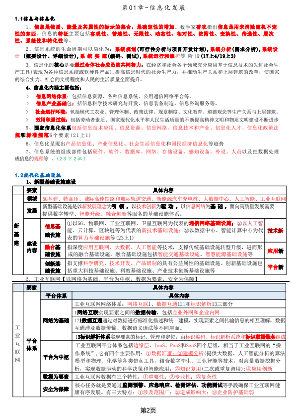 软考高项三色笔记
 镇安县双鑫矿业月河年处理15万吨尾渣综合加工利用项目水土保持报告表
  红杉资本:生成式AI最新市场格局.pdf
 wireshark 使用教程.pdf
 【2021年-贝佐斯致股东的信】.pdf
 巴菲特致股东的公开信 - 2022.pdf
 MySQL 8.1 参考手册.pdf
 世界银行报告下载:激进政策缩紧浪潮不足遏制通胀 全球经济衰退迫在眉睫(Is a Global Recession Imminent?).pdf
 红杉资本报告:适应与忍耐(Adapting to Endure).pdf
 高保真音响系统设计制作-毕业论文.doc
 一种自适应互补滤波姿态估计算法.pdf